Trending With Impact: Radiation, Senescence and Senotherapeutics

Researchers examined the effects of thoracic radiation-induced senescent cells on tumor progression, and the role of senotherapeutics to mitigate these effects.

Radiation therapy, advanced medical linear accelerator in therapeutic oncology to treat cancer

The Trending With Impact series highlights Aging (Aging-US) publications that attract higher visibility among readers around the world online, in the news, and on social media—beyond normal readership levels. Look for future science news about the latest trending publications here, and at Aging-US.com.

Listen to an audio version of this article

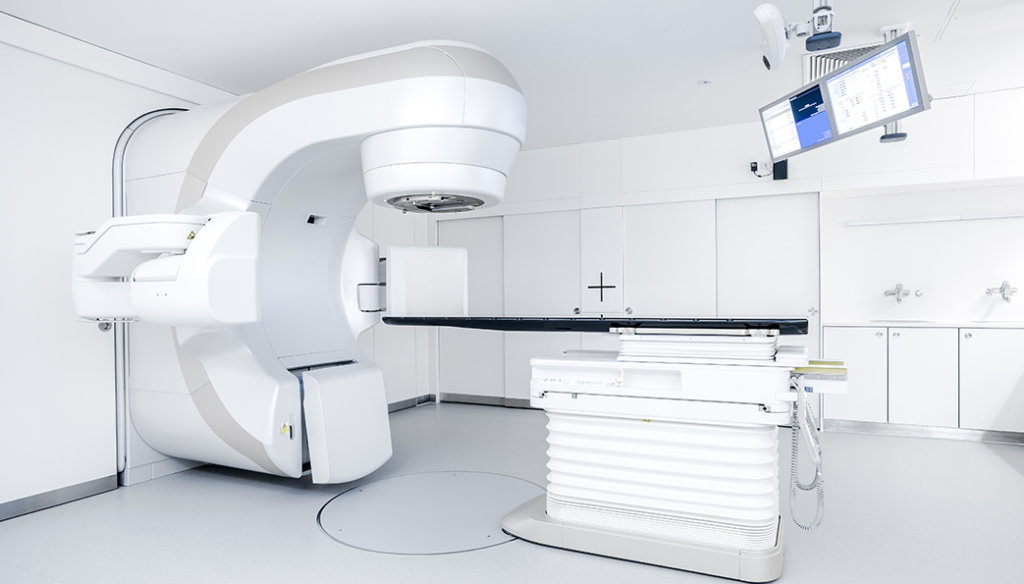
Radiation therapy is a highly-efficacious inducer of cancer cell death. With this being said, radiation has also previously been shown to cause premature senescence in the lung parenchyma. Senescence in cancer cells was previously only thought of as a mechanism capable of suppressing tumor cell proliferation by halting the cell cycle. However, a growing body of evidence shows that senescent cells may play a pro-tumorigenic role in cancer.

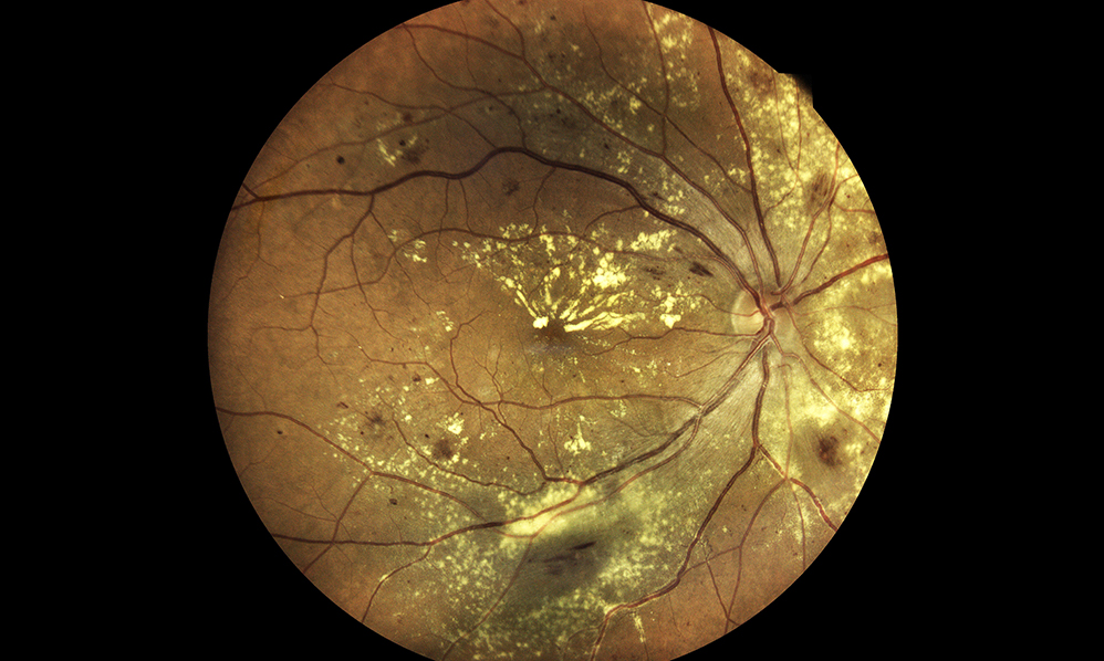
In the tumor microenvironment, the accumulation of senescent cells can become tumorigenic due to a lack of normal tissue stem cells and due to the expression of the senescence-associated secretory phenotype (SASP). SASP expression is when senescent cells secrete high levels of inflammatory cytokines, immune modulators, growth factors, and proteases. In addition to reinforcing senescence, SASP can create a biological environment that is immuno-suppressed and tumor-permissive. Radiation-induced senescence has previously been shown to have negative impacts on cancer patients.

“Cells that have undergone premature senescence due to stress, such as irradiation, are resistant to apoptotic cell death and effectively escape immune surveillance, resulting in their accumulation in tissue over time.”

Recently, researchers from the National Cancer Institute investigated the irradiated lung and the impact of radiation-induced senescent parenchymal cells on tumor growth. They also explored three senotherapeutics, rapamycin, INK-128 and ABT-737, for their potential to mitigate radiation-induced senescence. On February 12, 2022, the team’s priority research paper was published on the cover of Aging (Aging-US) Volume 14, Issue 3, and entitled, “Senescence-associated tumor growth is promoted by 12-Lipoxygenase.”

The Study

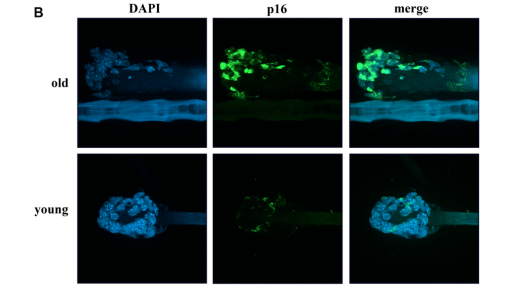
In this study, researchers intravenously injected melanoma cells into murine models two, four and eight weeks after daily fractions of thoracic irradiation exposure. There was also a control arm of unirradiated murine models. Tumor development was monitored by the number and size of the nodules in lung tissues. The number of cells exhibiting senescent activity was also recorded after two, four and eight weeks of thoracic irradiation. Their data demonstrated a correlation between the time points when tumors developed in the irradiated lungs and a marked accumulation of senescent cells.

“As previously described, in irradiated lungs, senescent cells increased significantly 4 and 8 weeks after IR compared to age matched unirradiated controls (Figure 1A).”

A characteristic of oncogene- and stress-induced senescence is the activation of mTOR signaling. Given this connection, the researchers conducted parallel studies evaluating senostatic agents capable of targeting the mTOR pathway, rapamycin and INK-128, and a senolytic agent to selectively eliminate senescent cells, ABT-737.  The data showed that rapamycin and INK-128 significantly reduced the number of tumor nodules in the lungs of irradiated mice compared to the controls. ABT-737 demonstrated reduced pulmonary senescence in irradiated mice.

The researchers also studied 12-Lipoxygensae (12-LOX), an enzyme that metabolizes a certain SASP molecule previously implicated in pulmonary senescence: 12(S)-HETE. 12-LOX is a known contributor to radiation-induced senescence and lung injury. The team specifically focused on the role of 12-LOX in pulmonary senescence and its impact on radiation-enhanced tumor growth. They found that inhibiting 12-LOX activity reduced radiation-induced lung senescence and mitigated radiation-enhanced tumor growth.

“Finally, we link senescence associated 12-LOX activity and production of 12(S)-HETE to the observed enhanced tumor growth after irradiation.”

Conclusion

In sum, the researchers found that radiation therapy can induce senescence in the lung parenchyma and also enhance tumor growth. The contribution of senescence in tumor progression was emphasized by the protection delivered by the mTOR-targeted senostatic and senolytic agents. This important discovery could lead to new therapies for cancer patients who are undergoing radiation therapy.

“Together, this study demonstrates the critical role of senescence in mediating radiation-enhanced tumor growth and identifies Alox12 as an important player in this phenomenon. Treatment with a senostatic agent, INK-128, identified in this study, or with agents like rapamycin and ABT-737 suggested their potential therapeutic use in alleviating radiation associated tumor growth.”

Click here to read the full priority research paper published by Aging (Aging-US).

AGING (AGING-US) VIDEOS: YouTube | LabTube | Aging-US.com

Aging (Aging-US) is an open-access journal that publishes research papers bi-monthly in all fields of aging research. These papers are available to read at no cost to readers on Aging-us.com. Open-access journals offer information that has the potential to benefit our societies from the inside out and may be shared with friends, neighbors, colleagues, and other researchers, far and wide.

For media inquiries, please contact media@impactjournals.com.

Trending With Impact: ARDD21 Meeting Report Highlights

Read a brief summary of a meeting report from the 8th Annual Aging Research and Drug Discovery (ARDD21) meeting. 

ARDD21

The Trending With Impact series highlights Aging (Aging-US) publications that attract higher visibility among readers around the world online, in the news, and on social media—beyond normal readership levels. Look for future science news about the latest trending publications here, and at Aging-US.com.

Listen to an audio version of this article

The 8th Annual Aging Research and Drug Discovery (ARDD21) meeting was held in Copenhagen, Denmark, from August 30 to September 3, 2021. This meeting was attended by over 130 people on-site, with an additional 1800 people engaged online. The focus of this meeting was the current landscape of aging research and various ways it can be applied to drug discovery. Topics included: age-dependent control of cellular maintenance processes, longevity pathways, artificial intelligence-based drug screening, cellular stress and aging, the benefits of dietary restriction, stem cell rejuvenation, senolytics as an aging therapeutic, diverse models of aging, aging clocks and biomarkers of aging, new ideas in preclinical and clinical aging research, the longevity industry landscape, and a Longevity Medicine Workshop.

In total, there were 75 presentations given at ARDD21 by prominent and dedicated aging researchers. The meeting was thoroughly summarized in a paper published in Aging (Aging-US) Volume 14, Issue 2, entitled, “Meeting Report: Aging Research and Drug Discovery.”

ARDD21 Meeting Report Highlights

One of the keynote presentations was given by Nir Barzilai from the Albert Einstein College of Medicine. He discussed his work on aging and how it can be applied to drug discovery. One interesting finding that he discussed was that many drugs currently used to treat chronic diseases, such as diabetes and heart disease, also have the potential to treat aging. This is due to the fact that many diseases are symptoms of aging, and thus, treating the underlying cause (aging) can in turn treat the symptoms.

Another keynote presentation was given by James Kirkland from the Mayo Clinic. He discussed his work on developing therapies to target senescent cells. Senescent cells can accumulate with age, and their presence has been linked with a variety of age-related conditions such as arthritis, cancer and heart disease. Kirkland’s team has developed a number of potential therapies to eliminate or reduce the number of senescent cells in the body, and he is currently testing them in clinical trials.

Professor Dame Linda Partridge from Max Planck Institute for Biology of Ageing presented on aging and the importance of intestinal homeostasis. Her studies involved rapamycin treatment to act on the longevity pathway mTOR, which revealed that short term and early treatment with rapamycin extends lifespan in D. melanogaster as much as chronic rapamycin treatment. Yu-Xuan Lu, another researcher from the Max Planck Institute for Biology of Ageing, demonstrated the existence of an unconventional intestine sex-specific TORC1-histone axis which uncovers a new aspect of improved longevity with rapamycin.

Brian Kennedy from the Buck Institute for Research on AgingNational University of Singapore and National University Health System showed how Alzheimer’s disease can be used as a model of neuronal aging. Presenting their new WormBot, Matt Kaeberlein from the University of Washington described a “set it and forget it” method of large-scale intervention testing in roundworms (C. elegans). 

“He stressed the importance of broad and unbiased screening of intervention beyond known pathways and in different combinations [56].”

Aging (Aging-US) Editorial Board member Alexey Moskalev from the Russian Academy of Sciences presented on the disruption of hydrogen sulfide homeostasis and its association with aging, and therefore, its potential as a gero-therapeutic target. David Sinclair from Harvard Medical School (also on the Aging Editorial Board) discussed aging-driven epigenetic and gene expression changes in the central nervous system. He showed that this can be safely reversed to restore vision by inducible adeno-associated viruses expressing polycistronic Oct4, Sox2 and Kif4, and that the effect is dependent on DNA demethylation. Finally, a Longevity Medicine Workshop was held with a panel of experts aimed to inspire young students to engage in longevity research. This panel included Aging Editorial Board members Alex Zhavoronkov, Alexey Moskalev and Mikhail Blagosklonny (Editor-In-Chief).

Conclusion

Overall, the ARDD21 meeting was a fruitful exhibition of experts from all areas of aging research that came together to share their latest findings in the field. The highlights in this blog pale in comparison to the thoughtful details included in the original meeting report. 

Click here to read the full meeting report published by Aging (Aging-US).

AGING (AGING-US) VIDEOS: YouTube | LabTube | Aging-US.com

Aging (Aging-US) is an open-access journal that publishes research papers bi-monthly in all fields of aging research. These papers are available to read at no cost to readers on Aging-us.com. Open-access journals offer information that has the potential to benefit our societies from the inside out and may be shared with friends, neighbors, colleagues, and other researchers, far and wide.

For media inquiries, please contact media@impactjournals.com.

How Habitual Tea Drinking Impacts Brain Structure

In 2019, researchers conducted the first study to explore the effects of habitual tea drinking on system-level brain networks.

Figure 3. Brain regions exhibiting significant differences in structural nodal efficiency between the tea drinking group and the non-tea drinking group at the significance level of 0.01 (uncorrected) statistical evaluated by a permutation test.
Listen to an audio version of this article

After water, tea is the most popular beverage in the world. While many people enjoy tea for the flavor, aroma and caffeine boost, research suggests that there may be another reason to regularly drink this beverage: its effects on the brain. In 2019, researchers from Wuyi UniversityUniversity of EssexUniversity of Cambridge, and the National University of Singapore conducted the first study exploring the effects of tea on system-level brain networks. Their paper was published in Aging (Aging-US) Volume 11, Issue 11, and entitled, “Habitual tea drinking modulates brain efficiency: evidence from brain connectivity evaluation.”

“In this study, we comprehensively explored brain connectivity with both global and regional metrics derived from structural and functional imaging to unveil putative differential connectivity organizations between tea drinking group and non-tea drinking group.”

The Study

The subjects enrolled in this study were older adults (mean ≈ 70 years old) from residential communities in Singapore, without conditions or terminal illnesses (see Materials and Methods). Researchers initially recruited 93 participants, however, only 36 total participants (male = 6; female = 30) remained after adjusting for the strict study inclusion criteria. Researchers classified the remaining participants as “non-tea drinkers” or “tea drinkers” using complex composite test scores. The composite score included self-reports of multiple decades of weekly green tea, oolong tea, black tea, and coffee intake (see Materials and Methods). After screening, 15 participants were assigned to the tea-drinking group and 21 were assigned to the non-tea drinking group. (Coffee intake did not differ significantly between the two groups.)

Next, structural brain connectivity was compared between tea drinkers and non-tea drinkers. All 36 participants underwent MRI brain scans and both functional and structural networks were investigated from global and regional perspectives. The researchers found that participants in the tea-drinking group had more efficient structural organization. However, tea did not seem to have a significantly beneficial effect on global functional organization. As a result of tea drinking, hemispheric asymmetry in the structural connectivity network was observed, although it was not observed in the functional connectivity network. 

“In addition, functional connectivity strength within the default mode network (DMN) was greater for the tea-drinking group, and coexistence of increasing and decreasing connective strengths was observed in the structural connectivity of the DMN.”

Conclusion

The researchers found that tea drinkers had more efficient brain structure organization than non-tea drinkers. Studies have previously demonstrated that tea drinkers are less likely to develop dementia, and tea consumption has also been linked with better cognitive performance. The researchers note that these effects are due to tea’s contents of caffeine, L-theanine and polyphenols (catechins). Polyphenols are compounds found in plants, including tea leaves, and may help protect against oxidative damage. Previous studies have shown that tea polyphenols can cross the blood-brain barrier and may help improve brain function. 

While this study’s findings suggest that habitual tea drinking leads to better brain connectivity and efficiency in old age, the researchers were forthcoming about the limitations of their study. The sample size was limited and other substances, behaviors, habits, and environmental factors may have impacted the outcome of the study. 

“Our study offers the first evidence of the positive contribution of tea drinking to brain structure and suggests a protective effect on age-related decline in brain organisation.”

Click here to read the full research paper published by Aging (Aging-US).

AGING (AGING-US) VIDEOS: YouTube | LabTube | Aging-US.com

Aging (Aging-US) is an open-access journal that publishes research papers monthly in all fields of aging research and other topics. These papers are available to read at no cost to readers on Aging-us.com. Open-access journals offer information that has the potential to benefit our societies from the inside out and may be shared with friends, neighbors, colleagues, and other researchers, far and wide.

For media inquiries, please contact media@impactjournals.com.

Aging’s Top 10 Most-Viewed Papers in 2021

Aging's Top 10 papers of 2021

Read the 10 most-viewed papers on Aging-US.com of 2021.

Listen to an audio version of this post

#10: Iron: an underrated factor in aging

Author: Dennis Mangan

Institution: MTOR LLC

Quote: “Blocking iron absorption through drugs or natural products extends lifespan. Many life-extending interventions, such as rapamycin, calorie restriction, and old plasma dilution can be explained by the effects they have on iron absorption, excretion, and metabolism.”


#9: Reversal of cognitive decline: A novel therapeutic program

Author: Dale E. Bredesen

Institutions: University of California Los Angeles and Buck Institute for Research on Aging

Quote: “This report describes a novel, comprehensive, and personalized therapeutic program that is based on the underlying pathogenesis of Alzheimer’s disease, and which involves multiple modalities designed to achieve metabolic enhancement for neurodegeneration (MEND).”


#8: Shorter telomere lengths in patients with severe COVID-19 disease

Authors: Raul Sanchez-Vazquez, Ana Guío-Carrión, Antonio Zapatero-Gaviria, Paula Martínez, and Maria A. Blasco

Institutions: Spanish National Cancer Research Center – CNIO and Field Hospital COVID-19, IFEMA

Quote: “The incidence of severe manifestations of COVID-19 increases with age with older patients showing the highest mortality, suggesting that molecular pathways underlying aging contribute to the severity of COVID-19. One mechanism of aging is the progressive shortening of telomeres, which are protective structures at chromosome ends.”


#7: Hyperbaric oxygen therapy alleviates vascular dysfunction and amyloid burden in an Alzheimer’s disease mouse model and in elderly patients

Authors: Ronit Shapira, Amos Gdalyahu, Irit Gottfried, Efrat Sasson, Amir Hadanny, Shai Efrati, Pablo Blinder, and Uri Ashery 

Institutions: Tel Aviv University and Assaf Harofeh Medical Center

Quote: “Hyperbaric oxygen therapy (HBOT) is in clinical use for a wide range of medical conditions. In the current study, we exposed 5XFAD mice, a well-studied AD model that presents impaired cognitive abilities, to HBOT and then investigated the therapeutical effects using two-photon live animal imaging, behavioral tasks, and biochemical and histological analysis.”


#6: Fighting the storm: could novel anti-TNFα and anti-IL-6 C. sativa cultivars tame cytokine storm in COVID-19?

Authors: Anna Kovalchuk, Bo Wang, Dongping Li, Rocio Rodriguez-Juarez, Slava Ilnytskyy, Igor Kovalchuk, and Olga Kovalchuk

Institutions: Pathway Research Inc.University of Calgary and University of Lethbridge

Quote: “Cannabis sativa has been proposed to modulate gene expression and inflammation and is under investigation for several potential therapeutic applications against autoinflammatory diseases and cancer. Here, we hypothesized that the extracts of novel C. sativa cultivars may be used to downregulate the expression of pro-inflammatory cytokines and pathways involved in inflammation and fibrosis.”


#5: Examining sleep deficiency and disturbance and their risk for incident dementia and all-cause mortality in older adults across 5 years in the United States

Authors: Rebecca Robbins, Stuart F. Quan, Matthew D. Weaver, Gregory Bormes, Laura K. Barger, and Charles A. Czeisler

Institutions: Brigham and Women’s HospitalHarvard Medical School and Boston College

Quote: “Sleep disturbance and deficiency are common among older adults and have been linked with dementia and all-cause mortality. Using nationally representative data, we examine the relationship between sleep disturbance and deficiency and their risk for incident dementia and all-cause mortality among older adults.”


#4: Rejuvant®, a potential life-extending compound formulation with alpha-ketoglutarate and vitamins, conferred an average 8 year reduction in biological aging, after an average of 7 months of use, in the TruAge DNA methylation test

Authors: Oleksandr Demidenko, Diogo Barardo, Valery Budovskii, Robb Finnemore, Francis R. Palmer III, Brian K. Kennedy, and Yelena V. Budovskaya

Institutions: TruMe Inc.National University SingaporePonce de Leon HealthNational University Health System Singapore, and Singapore Institute for Clinical Sciences, A*STAR

Quote: “Instead, aging biomarkers, such as DNA methylation (DNAm) clocks, have been developed to monitor biological age. Herein we report a retrospective analysis of DNA methylation age in 42 individuals taking Rejuvant®, an alpha-ketoglutarate based formulation, for an average period of 7 months.”


#3: Aging and rejuvenation – a modular epigenome model

Authors: Priscila Chiavellini, Martina Canatelli-Mallat, Marianne Lehmann, Maria D. Gallardo, Claudia B. Herenu, Jose L. Cordeiro, James Clement, and Rodolfo G. Goya

Institutions: National University of La PlataNational University of CordobaWorld Academy of Art and Science (WAAS), and Betterhumans Inc.

Quote: “The view of aging has evolved in parallel with the advances in biomedical sciences. Long considered as an irreversible process where interventions were only aimed at slowing down its progression, breakthrough discoveries like animal cloning and cell reprogramming have deeply changed our understanding of postnatal development, giving rise to the emerging view that the epigenome is the driver of aging.”


#2: Potential reversal of epigenetic age using a diet and lifestyle intervention: a pilot randomized clinical trial

Authors: Kara N. Fitzgerald, Romilly Hodges, Douglas Hanes, Emily Stack, David Cheishvili, Moshe Szyf, Janine Henkel, Melissa W. Twedt, Despina Giannopoulou, Josette Herdell, Sally Logan, and Ryan Bradley

Institutions: Institute for Functional MedicineAmerican Nutrition AssociationNational University of Natural MedicineAriel UniversityMcGill University, and University of California San Diego

Quote: “Manipulations to slow biological aging and extend healthspan are of interest given the societal and healthcare costs of our aging population. Herein we report on a randomized controlled clinical trial conducted among 43 healthy adult males between the ages of 50-72.”


#1: Hyperbaric oxygen therapy increases telomere length and decreases immunosenescence in isolated blood cells: a prospective trial

Authors: Yafit Hachmo, Amir Hadanny, Ramzia Abu Hamed, Malka Daniel-Kotovsky, Merav Catalogna, Gregory Fishlev, Erez Lang, Nir Polak, Keren Doenyas, Mony Friedman, Yonatan Zemel, Yair Bechor, and Shai Efrati

Institutions: Shamir Medical CenterTel Aviv University and Bar Ilan University

Quote: “At the cellular level, two key hallmarks of the aging process include telomere length (TL) shortening and cellular senescence. Repeated intermittent hyperoxic exposures, using certain hyperbaric oxygen therapy (HBOT) protocols, can induce regenerative effects which normally occur during hypoxia. The aim of the current study was to evaluate whether HBOT affects TL and senescent cell concentrations in a normal, non-pathological, aging adult population.”


Click here to read the latest papers published by Aging.

AGING (AGING-US) VIDEOS: YouTube | LabTube | Aging-US.com

Aging (Aging-US) is an open-access journal that publishes research papers bi-monthly in all fields of aging research. These papers are available to read at no cost to readers on Aging-us.com. Open-access journals offer information that has the potential to benefit our societies from the inside out and may be shared with friends, neighbors, colleagues, and other researchers, far and wide.

For media inquiries, please contact media@impactjournals.com.

Trending With Impact: Are Our Muscles Intrinsically Impaired by Aging?

In a priority research paper published by Aging-US in January of 2022, researchers investigated aged muscle stem cells and their ability to sense and respond to mechanical cues.

Listen to an audio version of this article
3D Illustration of muscle tissue

The Trending With Impact series highlights Aging (Aging-US) publications that attract higher visibility among readers around the world online, in the news, and on social media—beyond normal readership levels. Look for future science news about the latest trending publications here, and at Aging-US.com.

IIs muscle wasting a fate humans can avoid, or will the problem of aging-related muscle loss only be resolved when the mystery of aging is solved? Researchers—from Vrije Universiteit AmsterdamUniversity of AmsterdamSorbonne UniversitéAmsterdam University Medical Center VUmcUniversité Catholique de LouvainKU Leuven, and Institut NeuroMyoGène—conducted a study aimed at elucidating whether muscle stem cells are inherently impaired by the aging process in their ability to sense and respond to mechanical cues. Their priority research paper was published in January of 2022 on the cover of Aging (Aging-US) Volume 14, Issue 1, and entitled, “Reduced growth rate of aged muscle stem cells is associated with impaired mechanosensitivity.”

Muscle Stem Cells

Muscle stem cells (MuSCs) are stem cells located within skeletal muscle tissues. MuSCs function to repair Muscle stem cells (MuSCs) are stem cells located within skeletal muscle tissues. MuSCs function to repair damaged myofibers and give rise to new skeletal muscle cells. These self-renewing stem cells are involved in muscle growth, repair and regeneration. As we age, MuSCs decline in number and lose their potential to regenerate damaged myofibers, leading to sarcopenia. The researchers in this study hypothesized that the responsiveness of aged MuSCs is impared by the aging process both physically and mechanically.

“We postulated that aged MuSCs are intrinsically impaired in their responsiveness to omnipresent mechanical cues through alterations in MuSC morphology, mechanical properties, and number of integrins, culminating in impaired proliferative capacity.”

The Study

The researchers assessed whether aged MuSCs become impaired in their ability to proliferate, respond to pulsating fluid shear stress (PFSS) mechanical loading, maintain focal adhesion number and/or size after mechanical loading, and in their ability to express the protein-coding gene Integrin Subunit Alpha 7 (ITGA7). 

“Integrins are transmembrane protein receptors that connect MuSCs to the ECM [extracellular matrix] components and are part of focal adhesions [51].”

Young MuSCs (2 months) and aged MuSCs (22 months) were isolated from male mice. Fluorescence-activated cell purification was carried out and cells were cultured. To measure proliferation, images were captured of the cell cultures every 24 hours. Images were also taken pre- and post-PFSS to determine the number of young and aged MuSCs detached from the culture media (focal adhesion) as a result of PFSS treatment. Since nitric oxide (NO) is known to play a role in MuSC activation and muscle regeneration, NO analysis was conducted to measure NO production. To determine MuSC morphology, the researchers carried out immunohistochemistry staining. They also measured MuSC stiffness, deformation, gene expression, and RNA isolation and reverse transcription.

Compared to young MuSCs, the researchers found aged MuSCs had impaired growth. Their results showed that IL-6 gene expression was lower in aged MuSCs, which suggested that aged MuSCs were intrinsically altered in the signaling pathways governing proliferation and MuSC function. Aged MuSCs showed an increase in cell volume and reduced cell adhesion after mechanical loading. NO levels in young and aged MuSCs were similar, and PFSS in both cultures resulted in similar increases in NO production. The researchers found decreased ITGA7 expression and reduced pPXN clusters (focal adhesion formation) were involved in altered MuSC function with age. High YAP nuclear localization was found in aged MuSCs, as well as reduced mechanosensitivity.

“Aged MuSCs were less sensitive to shear forces and showed upregulation of less genes, suggesting that the decreased mechanosensitivity was due to decreased integrin protein expression, i.e. ITGA7, ITGA5, and ITGB5, and focal adhesion number.”

Conclusion

The results from this study found that aged MuSCs were intrinsically impaired in their growth rate due to decreased ITGA7 expression and diminished focal adhesion formation. These changes coincided with increased cell volume, decreased MuSC adhesion, altered mechanosensitivity, changed YAP signaling and decreased expression of several genes (including cell cycle genes). The researchers suggest that ITGA7 and pPXN may be potential therapeutic targets to improve aged MuSC function.

“As an implication, a possible therapeutic option could be restoration ITGA7 and focal adhesion number in aged MuSCs, which may help to restore MuSCs adhesion to their niche as well as growth rate of these cells.”

Click here to read the full priority research paper published by Aging (Aging-US).

AGING (AGING-US) VIDEOS: YouTube | LabTube | Aging-US.com

Aging (Aging-US) is an open-access journal that publishes research papers bi-monthly in all fields of aging research. These papers are available to read at no cost to readers on Aging-us.com. Open-access journals offer information that has the potential to benefit our societies from the inside out and may be shared with friends, neighbors, colleagues, and other researchers, far and wide.

For media inquiries, please contact media@impactjournals.com.

Common Age-Related Changes in Eye Lenses

In this 2019 study, researchers examined murine models to determine common age-related eye lens changes that contribute to eventual vision impairment and loss.

(Truncated) Figure 7. Whole lens staining for F-actin (phalloidin, green) and nuclei (DAPI, red) in 4-month-old and 18-month-old lenses.
Figure 7. Whole lens staining for F-actin (phalloidin, green) and nuclei (DAPI, red) in 4-month-old and 18-month-old lenses. (Truncated)
Listen to an audio version of this article

A variety of eye disorders can occur as humans age, including age-related macular degeneration, cataracts, presbyopia, glaucomadry eyes, and temporal arteritis. These conditions can contribute to vision impairment and even vision loss. Unfortunately, the full gambit of common age-related eye lens changes which contribute to these disorders is not yet fully defined. However, while mice and primates are different species, their eye lenses share common characteristics. This means that studies in murine models regarding age-related eye lens changes may provide a baseline for aging studies on human eye lenses in the future. 

“Little is known about the morphological, mechanical, refractive and cellular changes that occur with advanced age in the lens. Mice offer an opportunity to investigate changes in lens morphometrics, stiffness, transparency and refractive properties with age in a relatively shortened period of time.”

To further define common age-related changes in eye lenses, researchers—from The Scripps Research InstituteUniversity of DelawareMorehouse School of MedicineNottingham Trent UniversityJapan Synchrotron Radiation Research Institute, and Boston University School of Medicine—conducted an extensive study of eye lenses among mice between one and 30 months of age. Their paper was published by Aging (Aging-US) in 2019, and entitled, “Age-related changes in eye lens biomechanics, morphology, refractive index and transparency.”

The Study

In this study, the researchers measured the size, refractive index (Gradient Refractive Index, GRIN) and stiffness of mouse lenses in young adult mice, starting at one and two months old, to very old mice of 24 to 30 months old. The team examined mechanisms of age-related cataracts, cell morphology in aged lenses, increased lens stiffness with age, and lens resilience. Methods used in this study include: lens biomechanical testing and morphometrics, live lens imaging, capsule thickness and fiber cell width measurements, phalloidin-staining of epithelial cells in whole lenses, scanning electron microscopy, transmission electron microscopy, and X-ray talbot interferometry. 

The researchers found that, with age, mouse eye lenses increased in size, nuclear fraction, stiffness, and resilience. After four months of age, lens capsule thickness and fiber cell width did not increase, but epithelial cell area increased slightly with age. In the lenses of mice older than 12 months, the researchers observed anterior cataracts, cortical haziness and ring cataracts. They found that the anterior cataracts were due to incomplete suture closure and detachment of anterior epithelial cells from the underlying fiber cells. The ring cataracts were linked to abnormal compaction of differentiating fiber cells. The hexagonal packing of fiber cells was shown to be disrupted with age. Lastly, the researchers observed that the gradient refractive index increased and then plateaued with age.

“Our comprehensive study of aging in wild-type mouse lenses in the B6 genetic background showed increased stiffness along with appearance of anterior, cortical and ring cataracts with age (Figure 14).”

Conclusion

Overall, the researchers demonstrate that age-related changes in mouse lenses mimic some aspects of aging in human lenses. Aside from the obvious study limitations (mouse-to-human translation), the data collected from this study provide a comprehensive overview of age-related changes in murine lenses, including lens size, stiffness, nuclear fraction, refractive index, transparency, capsule thickness, and cell structure.

“Whether there is a common molecular mechanism that drives changes in all the measured parameters remains unknown, but further biochemical and cell morphology studies will be needed to determine how subcellular aging affects the whole tissue.”

Click here to read the full research paper published by Aging (Aging-US).

AGING (AGING-US) VIDEOS: YouTube | LabTube | Aging-US.com

Aging (Aging-US) is an open-access journal that publishes research papers bi-monthly in all fields of aging research and other topics. These papers are available to read at no cost to readers on Aging-us.com. Open-access journals offer information that has the potential to benefit our societies from the inside out and may be shared with friends, neighbors, colleagues, and other researchers, far and wide.

For media inquiries, please contact media@impactjournals.com.

Aging and Circadian Rhythm: Does a Conserved Link Exist?

In the Aging (Aging-US) Volume 13, Issue 24, cover paper, researchers conducted a study suggesting that the circadian rhythm is subjected to aging-related gene alterations.

Figure 6. Matching of our CR-related DEGs evidenced to be regulated with aging with a curated human CR network.
Listen to an audio version of this article

Chronobiology is the study of biological rhythms. The human circadian system is a biological process known to regulate the sleeping and waking cycle (circadian rhythm; CR). Components of the circadian system are known as ​​clock genes. Clock genes generate daily oscillations of gene expression and interact as an intricate network to influence biological processes in organisms, tissues and cells. This system is primarily regulated by Earth’s day and night cycles (light and darkness), though it can be affected by other factors, including nutrition, cellular devices, stress, illness, jet lag, and aging.

“It is well established that aging interferes with the regulation of the circadian system, which, in return, contributes to the manifestation and progression of aging-related diseases (reviewed in [45]).”

Across an organism’s lifespan, changes in circadian rhythm take place. These changes can cause aging-related diseases to become more prevalent. Studies have also shown that age-independent alterations to the circadian system can result in premature aging. This interrelation between aging and CR means that aging may play a role in the circadian system and that the circadian system may play a role in aging. However, researchers have not yet fully illuminated the impact of aging-related circadian system changes on healthy organs and tissues. 

“Whether aging-related changes of the circadian system’s regulation follow a conserved pattern across different species and tissues, hence representing a common driving force of aging, is unclear.”

The Study

In an effort to identify circadian rhythm regulatory patterns over the course of aging, researchers—from Friedrich Schiller University JenaFLI Leibniz Institute for Age ResearchJena University HospitalGerman Center for Integrative Biodiversity Research, and European Virus Bioinformatics Center—performed inter-species and inter-organ transcriptional analyses. The research paper was published in December of 2021 as the cover of Aging (Aging-US) Volume 12, Issue 24, and entitled, “Age-dependent expression changes of circadian system-related genes reveal a potentially conserved link to aging.”

“Here, we used RNA-Seq data to profile the regulation of CR-related genes of 4 different species in a cross-sectional study in individuals ranging from young mature to old-age categories.”

In this cross-sectional study, the researchers used data from 329 RNA sequencing libraries to identify differentially expressed genes in transcriptional profiles among humans, house mice, zebrafish, and the extremely short-lived turquoise killifish. All human donors were classified into the following age groups of 14 to 15 individuals: mature (24–29 years), aged (60–65 years), and old-age (75–79 years). The other species were categorized into their respective age groups. Organs, including the brain, blood, liver, and skin, were examined and then compared between the four species.

The researchers found that two circadian rhythm-related genes (dec2 and per2) were altered in all four species, primarily in early- and late-aging groups. Four genes (cirp, klf10, nfil3, and dbp) with aging-related expression patterns were found in several organs and species. In total, the researchers identified six genes (in several tissues from at least three out of the four species) that function at all regulation levels of circadian rhythm with apparently conserved age-associated regulation.

Conclusion

“​​Thus, these genes might represent a conserved link between the circadian system and aging.”

This study confirms work from previous studies and extends them by providing a new dataset linking circadian rhythm factors to physiological aging across four evolutionarily distinct species. Whether circadian rhythm regulation is the cause or a consequence of the aging process still remains to be explored. The researchers note that their non-synchronized cross-sectional approach should be replicated in the future and include an additional dataset based on a longitudinal study design, tissue synchronizations across species of interest and to potentially analyze anatomic sub-regions of the brain.

“In summary, our results show that modulations in CR-related gene transcription throughout aging are a conserved trait that is traceable across evolutionarily diverse species, ranging from humans to mice and fish.”

Click here to read the full research paper published by Aging (Aging-US).

AGING (AGING-US) VIDEOS: YouTube | LabTube | Aging-US.com

Aging (Aging-US) is an open-access journal that publishes research papers bi-monthly in all fields of aging research and other topics. These papers are available to read at no cost to readers on Aging-us.com. Open-access journals offer information that has the potential to benefit our societies from the inside out and may be shared with friends, neighbors, colleagues, and other researchers, far and wide.

For media inquiries, please contact media@impactjournals.com.

Investigating a Biomarker of Age-Related Macular Degeneration

Age-related macular degeneration
Ocular drusen in age-related macular degeneration (AMD).

Over 15 million people in the United States are currently struggling with age-related macular degeneration (AMD). Among aging populations around the world, AMD is the leading cause of irreversible vision loss. A main target of AMD is the pigmented layer of the retina, or the retinal pigment epithelium (RPE). The subretinal space of the eye encompasses the area under the retina between RPE cells and photoreceptors. Yellow deposits of lipids and proteins, called drusen, are located in the subretinal space and the hallmark sign of AMD. In 2005, researchers found that drusen deposits also contain an immune protein called the C-reactive protein (CRP).

Circulating CRP is one of the main clinical biomarkers of inflammation and infection, since CRP levels rise and fall with inflammation. This immune-response protein can exert pro-inflammatory properties by disassociating from its pentameric form (pCRP) into smaller monomeric CRP (mCRP) subunits. These mCRPs are small enough to cross the ocular blood–retinal barrier (oBRB) and appear in ocular drusen.

“mCRP has been identified in ocular drusen and other subepithelial deposits [2425], as well as in the choroid, and we have shown that mCRP, but not pCRP, contributes to oBRB disruption in vitro [26].”

Researchers—from Hospital Clínic de BarcelonaManchester Metropolitan UniversityHospital de la Santa Creu i Sant PauInstitute Salud Carlos IIIUniversity of BristolMoorfields Eye Hospital, and University College London Institute of Ophthalmology—conducted a study aimed at understanding the mCRP’s contribution to the pathophysiology of AMD. Their paper was published by Aging (Aging-US) in 2020, and entitled, “Activation of C-reactive protein proinflammatory phenotype in the blood retinal barrier in vitro: implications for age-related macular degeneration”.

The Study

“If mCRP pro-inflammatory capacity is unrestrained in AMD and particularly in high risk patients, then we need to determine how mCRP is generated or accumulates in the subretinal space as there is no CRP transcription in the retinal tissue [3031].”

In order to investigate how mCRP is generated and/or how it accumulates within the subretinal space, the researchers used a Transwell model to first determine whether circulating CRP could reach the subretinal space. The Transwell model included monolayers of primary porcine choroidal endothelial cells (CECs) grown on porous filters with their apical and basolateral surfaces exposed to separate chambers. They found that CRP isoforms were able to cross the CEC monolayer on the apical side of the RPE. Next, they used the Transwell model to evaluate whether CRP isoforms could also reach the subretinal space and cross RPE ARPE-19 cells. They found that mCRP was able to diffuse into both the subretinal space and cross into the RPE. pCRP was not found in the opposite chambers, even after 24 and 48 hours post-exposure. However, when the researchers tested younger and healthier primary porcine RPE cells instead of the ARPE-19 cells, neither pCRP nor mCRP diffused into opposite chambers.

Next, the researchers studied whether pCRP could dissociate into mCRP within the RPE. They found that induced inflammation triggered pCRP dissociation into mCRP in both the ARPE-19 and primary porcine RPE cells. The team also found that barrier disruption induced by mCRP was dependent on its topological localization.

Conclusion

“In summary, our findings further support mCRP direct contribution to progression of AMD, at least at the RPE level. The topological experiments elicit that mCRP is proinflammatory when present on the apical side of the RPE. However, mCRP is likely to only reach the apical side of the RPE in compromised RPE health and where barrier functions are compromised.”

The researchers were forthcoming about limitations in their study. Nonetheless, this study suggests that a plausible mechanism by which mCRP may contribute to RPE dysfunction and AMD progression is, when pCRP reaches the oBRB, it diffuses past the oBRB by dissociating into mCRP. It is also possible that mCRP may be derived from the dissociation of pCRP on the surface of damaged RPE. The proinflammatory microenvironment may be amplified and the barrier disruption may be enhanced when mCRP reaches the apical side of an already aged or damaged RPE.

“With respect to previous findings, this pathologic mechanism will be more prevalent in patients carrying the FH risk polymorphism for AMD, where mCRP proinflammatory effects remain unrestrained [28].”

Click here to read the full research paper published by Aging (Aging-US).

AGING (AGING-US) VIDEOS: YouTube | LabTube | Aging-US.com

Aging (Aging-US) is an open-access journal that publishes research papers bi-monthly in all fields of aging research and other topics. These papers are available to read at no cost to readers on Aging-us.com. Open-access journals offer information that has the potential to benefit our societies from the inside out and may be shared with friends, neighbors, colleagues, and other researchers, far and wide.

For media inquiries, please contact media@impactjournals.com.

Trending With Impact: Hair Follicles May Replace Traditional Biopsies

A new device has been developed by researchers to efficiently and painlessly collect hair follicle tissue samples from laboratory mammals, and even humans.

Figure 6. Markers of senescence analysis in hair follicular cells.

The Trending With Impact series highlights Aging (Aging-US) publications that attract higher visibility among readers around the world online, in the news, and on social media—beyond normal readership levels. Look for future science news about the latest trending publications here, and at Aging-US.com.

Laboratory mammals have impacted human-kind far beyond enhancing scientific knowledge in behavioral and environmental research. These animals have greatly contributed to human healthspan and lifespan in countless ways; from validating life-saving cancer therapies to accelerating the future of human anti-aging and longevity interventions. With respect for these salubrious animals, ethical standards (per country) require that researchers handle laboratory mammals with care, and that pain and stress are minimized. Blood and skin tissue samples (biopsies) collected from animals should be replaced whenever possible. For the researchers, this twofold invasive procedure for the animals is also time- and resource-limiting—presenting a bottleneck in the biomedical research process.

“However, we present here a simple method for obtaining biological material in the form of follicular cells from laboratory mice with sufficient quantities and quality for multiple analyses using standard modern molecular biology methods.”

In an effort to efficiently and humanely solve this ethics/logistics problem, researchers—from Palacky UniversityUniversity Hospital OlomoucDanish Cancer Society Research Center, and Karolinska Institute—developed a novel, non-invasive device that can be used to collect tissue samples from hair follicles. They tested the applications of this device and authored a research paper of the study. In December of 2021, their paper was published on the cover of Aging (Aging-US) Volume 13, Issue 23, and entitled, “An efficient, non-invasive approach for in-vivo sampling of hair follicles: design and applications in monitoring DNA damage and aging.”

“As millions of laboratory mice are routinely genotyped globally every year this approach represents a major ethical and logistic breakthrough.”

The Follicular Cells’ Collector

As opposed to traditional biopsies, hair follicle collection is a humane, easy, non-invasive, and painless method of DNA and tissue sample collection. Each hair follicle contains approximately 50 cells—of various cell types. 

“This micro-organ structure also has other advantages in biomarker studies, including suitability for investigations of circadian rhythms [57], and the presence of numerous cell types in a small area, which can be easily distinguished, such as keratinocytes, melanocytes, or perifollicular macrophages and mast cells [810].”

Previously, the limitations of using hair follicles as DNA and tissue samples stemmed from ineffective technology. Many devices involved ordinary tweezers and forceps with high risks for cross-contamination. The researchers termed their novel tissue sample collection device the “follicular cells’ collector.” The follicular cells’ collector is designed with dual pipettes and utilizes a precision vacuum method of hair follicle extraction. The device can be used to comfortably collect DNA and tissue samples from laboratory mammals, and even from humans. 

“Although hair samples have been previously used for that purpose [2931], our sample collection approach may motivate researchers to use them more routinely and widely.”

The Study

To validate that these hair follicle samples contain the required genetic information necessary in most studies, researchers compared murine genotyping results of 151 tail biopsies and 151 hair samples. In order to determine the ability of these samples to detect changes in expression patterns induced by external factors, the team also observed the DNA damage response in hair follicle cells after gamma irradiation and after the topical application of chemical clastogens. Further exploring its potential application in aging research, researchers assayed expression patterns of selected markers of biological age and senescence in murine hair follicular cells. The researchers conducted many other tests and experiments using murine hair follicular cells in this study.

“The speed by which the samples can be collected and processed (e.g. by fixation) is among the biggest advantages of our solution as it can be performed within seconds. This fact limits any potential underlying cellular responses and additional DDR [DNA damage response] caused by cofounding stressing factors related to the withdrawal process [2].”

Conclusion

The researchers found that the follicular cells’ collector method of obtaining mouse hair follicular cells can be successfully used for genotyping, quantitative polymerase chain reaction testing and quantitative immunofluorescence. They also demonstrated that this method can successfully monitor quality and expression level changes of selected proteins—induced by external factors and during natural or experimentally induced aging. 

“Our results highlight the value of hair follicles as biological material for convenient in vivo sampling and processing in both translational research and routine applications, with a broad range of ethical and logistic advantages over currently used biopsy-based approaches.”

Click here to read the full research paper published by Aging (Aging-US).

AGING (AGING-US) VIDEOS: YouTube | LabTube | Aging-US.com

Aging (Aging-US) is an open-access journal that publishes research papers bi-monthly in all fields of aging research and other topics. These papers are available to read at no cost to readers on Aging-us.com. Open-access journals offer information that has the potential to benefit our societies from the inside out and may be shared with friends, neighbors, colleagues, and other researchers, far and wide.

For media inquiries, please contact media@impactjournals.com.

Trending With Impact: Aging Reduced by 8 Years With Rejuvant®

A recent study revealed that participants experienced an average 8 year reduction in biological aging after taking Rejuvant® for approximately 7 months.

Anti-aging hourglass

The Trending With Impact series highlights Aging (Aging-US) publications that attract higher visibility among readers around the world online, in the news, and on social media—beyond normal readership levels. Look for future science news about the latest trending publications here, and at Aging-US.com.

Listen to an audio version of this article

Waiting until the end of a subject’s lifespan is quite a leaden method of validating the efficacy of a longevity-based intervention. This method could take researchers generations upon generations to eventually validate an effective intervention—or—this method might not ever yield results seen by the general public. However, researchers may have devised an innovative way to solve this problem.

“If we hope to control the aging process, we need to learn how to measure the rate of aging in shorter time periods.”

Many researchers believe that measuring the rate of human aging can be done faster by using DNA methylation-based aging clocks. Methylation-based clocks are capable of determining human biological aging with impressive accuracy. Hypermethylated and demethylated regions of DNA (CpG islands near specific aging-associated genes) play a key role in turning certain genes on and off throughout the aging process. Therefore, methylation is a biomarker of aging. While there is a short list of currently available biological aging clocks for researchers to use in studies of anti-aging therapies, the TruAge DNA methylation test is preferable in some cases, due to its accessibility, use of simple saliva samples and cost effectiveness.

“For the first time, these biomarkers of aging give scientists the opportunity to study the effects of anti-aging compounds in real-time and directly in humans.”

In a new study, researchers from TruMe LabsNational University of Singapore and Ponce de Leon Health used the TruAge DNA methylation test to validate Rejuvant®—a patent-pending anti-aging dietary supplement. The trial study yielded unprecedented results and the research paper authored by the team was published as the cover of Aging (Aging-US) Volume 13, Issue 22, entitled: “Rejuvant®, a potential life-extending compound formulation with alpha-ketoglutarate and vitamins, conferred an average 8 year reduction in biological aging, after an average of 7 months of use, in the TruAge DNA methylation test”.

The Study

Developed at Ponce de Leon Health, Rejuvant® is composed primarily of a compound called Alpha-Ketoglutarate (AKG). This molecule is naturally produced in humans and functions as a signaling molecule, an energy donor, a precursor to amino acid biosynthesis, and a regulator of epigenetic processes. In humans and other animals, AKG levels gradually decrease with age. Other components of Rejuvant® are calcium and, for males and females, Vitamin A and Vitamin D, respectively. 

“The goal of the study was to determine the effect of Rejuvant® supplementation on human biological aging by measuring DNA methylation.”

Researchers enrolled 42 healthy participants who were on average 64 years of age (43 to 72). Before taking Rejuvant®, all 42 participants completed a survey and their baseline biological age was measured using the TruMe age prediction model. The survey was a self-reported questionnaire including information about diet, alcohol intake, previous consumption of Rejuvant®, health, height and weight, sleep duration, smoking status, exercise frequency, physical activity level, meal frequency, snacking frequency, number of additional dietary supplements consumed and frequency, hair status, education, healthy lifestyle mindset, and trust in dietary supplements. 

Participants (majority male; 28) took two tablets of Rejuvant® daily, for a duration of four to 10 months. Biological age was measured from saliva samples again after taking Rejuvant® for four to 10 months. At the end of the trial, participants completed the same survey. The researchers compared the baseline surveys with the final surveys to check for other confounders contributing to the results in the study. They also used the surveys to select a sub-group of 13 participants who reported no change in diet type, drinking frequency, additional dietary supplements intake, sleep duration, and exercise frequency. They compared this sup-group with the rest of the cohort. They also compared results between males and females, older and younger participants, and participants with higher biological age relative to their chronological age (aging more quickly).

Results and Conclusion

Researchers examined associations between the epigenetic clock, health status, physical fitness, and the effects of Rejuvant® on human biological aging. The researchers were forthcoming about limitations in this study. A control arm was not used, the cohort was relatively small, only one biological aging clock was used, and researchers did not collect other kinds of data relevant to aging. However, the study results showed that Rejuvant® conferred an average eight year reduction in biological aging after approximately seven months of use. The 13 participants in the sub-group saw anti-aging benefits slightly less than the rest of the cohort. Rejuvant® was more effective in chronologically older participants and in participants that were aging more quickly (with a higher biological age relative to their chronological age).

“Future randomized clinical trials will be required to confirm the findings presented here. Nevertheless, the results in this manuscript suggest that Rejuvant® may have significant effects on biological age as measured by DNA methylation of saliva samples.”

Click here to read the full priority research paper published by Aging (Aging-US).

WATCH: AGING VIDEOS ON LABTUBE

Aging (Aging-US) is an open-access journal that publishes research papers monthly in all fields of aging research and other topics. These papers are available to read at no cost to readers on Aging-us.com. Open-access journals offer information that has the potential to benefit our societies from the inside out and may be shared with friends, neighbors, colleagues, and other researchers, far and wide.

For media inquiries, please contact media@impactjournals.com.

  • Follow Us