Is Estrogen Dysregulation Behind Alzheimer’s Pathology?

In a new study, researchers explored Alzheimer’s disease and its potential relationship with the estrogen receptor-α gene (ESR1).

In a new study, researchers explored Alzheimer's disease and its potential relationship with the estrogen receptor-α gene (ESR1).

The Trending With Impact series highlights Aging publications (listed as “Aging (Albany NY)” by Medline/PubMed and “Aging-US” by Web of Science) that attract higher visibility among readers around the world online, in the news, and on social media—beyond normal readership levels. Look for future science news about the latest trending publications here, and at Aging-US.com.

Listen to an audio version of this article

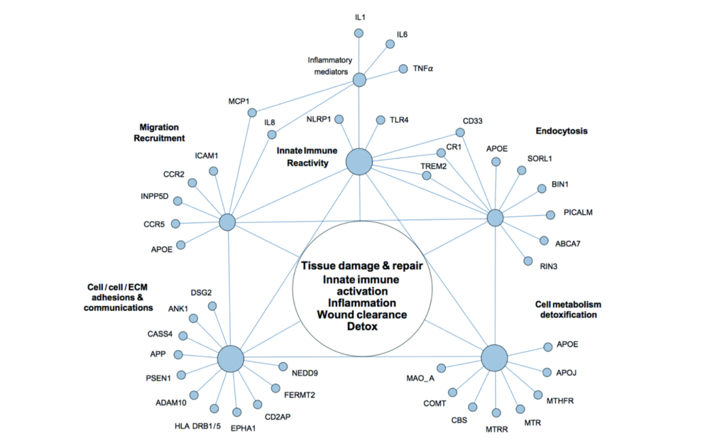
The United States government currently has a mind-blowing annual budget of $3.5 billion designated for Alzheimer’s disease (AD) and dementia research funding. Therapeutics pushed forward thus far have been largely based on the amyloid-beta (Aβ) cascade hypothesis of AD. Surprisingly, despite decades and billions, these interventions have yielded little to no benefits for AD patients. This lack of efficacy has encouraged some researchers to rethink AD pathology and focus on discovering key triggers and mechanisms of neuroinflammation.

“There has been a lengthy and ongoing scientific debate around the causative factors of AD, and the relative importance of both senile Aβ plaques and tau tangles has been largely informed by postmortem investigations of the AD brain. For several decades, the amyloid hypothesis has dominated the field, which has brought forth many high-profile therapeutic attempts that have produced side effects but no real benefits [5].”

Women & Alzheimer’s Disease

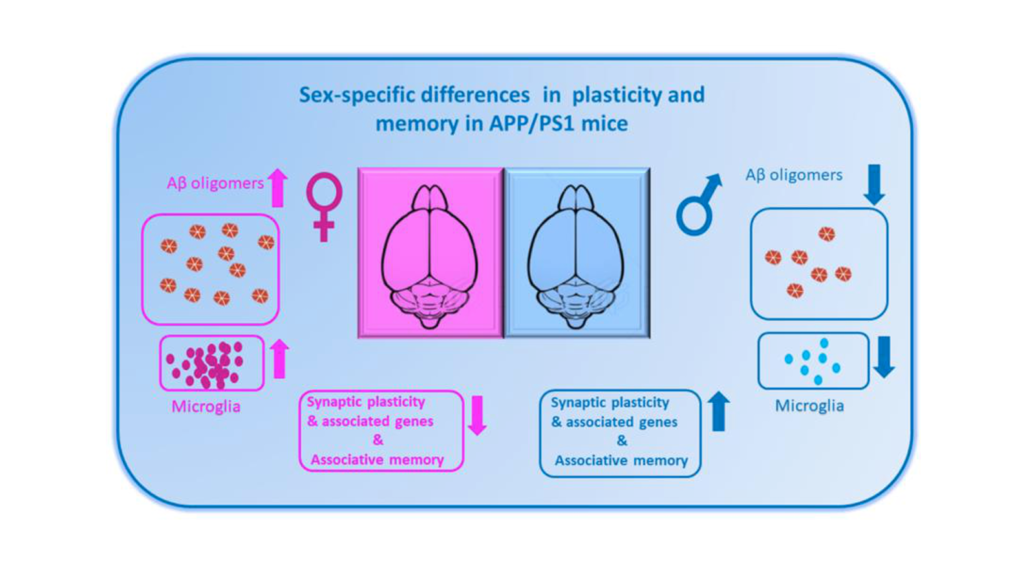
Women compose two-thirds of the United States Alzheimer’s population. Is this gender-specific risk a result of living longer or is it due to other causes, perhaps related to hormonal differences or gender-associated differential gene expression? Previous studies have found that estrogen may protect neurons from the damaging effects of amyloid-beta plaques and tau tangles. However, in women, estrogen levels tend to decline with age, which could be one reason why aging women are more susceptible to AD. 

In a new study, researchers Junying Liu, Shouli Yuan, Xinhui Niu, Robbie Kelleher, and Helen Sheridan from Trinity College Dublin, Peking University and Jilin University examined the potential relationship between the estrogen receptor-α gene (ESR1) and neuroinflammation. Their research paper was published on November 1, 2022, in Aging’s Volume 14, Issue 21, and entitled, “ESR1 dysfunction triggers neuroinflammation as a critical upstream causative factor of the Alzheimer’s disease process.”

“AD is characterized by three major questions: Why is age the primary risk factor? Why are women more sensitive to the onset of this form of dementia? And why are neurons in areas of the brain that are essential for memory selectively targeted?”

The Study

Originally, the researchers in this study had been in the process of investigating ESR1-knockdown in breast cancer when they stumbled upon another discovery. (ESR1 is a gene that codes for the estrogen receptor, a protein that helps to regulate cell division and differentiation.) To their surprise, KEGG pathway enrichment analysis showed that ESR1 may also be related to axonal guidance, inflammation-related gene markers and Notch signaling pathways. Upon further validation using a dataset of in vivo AD inflammatory samples, the team found that the ESR1 gene was altered in AD patients and was associated with an increase in pro-inflammatory markers.

“ESR dysfunction likely plays a role in AD pathology – especially in women – although the specific mechanisms remain unclear. In vivo and ex vivo studies demonstrate that neuroinflammatory brain states overlap with ESR signaling pathways and that these two systems interact closely.”

In the current study, the researchers used an animal model to explore the potential role of ESR1 in modulating inflammation-related AD pathology. Using a macrophage cell line, they identified ESR1 as a key modulator of inflammation in the context of AD. They then showed that when the ESR1 gene was absent or mutated, neuroinflammation occurred. This finding offers a potential mechanism for understanding the gender-specific risk of AD in women.

“Our results suggest that ESR1 is modulated by apolipoprotein E (APOE) through CEBPB/ATF4, mir-155-5p, or mir-1-3p. Moreover, sea hare-hydrolysates (SHH), as one of the axonal guidance molecules, could regulate the STAT3/PRDM1/CEBPB pathway and consequently induce cell death through pyroptosis signaling pathways, trigger the secretion of IL1β, leading to neuroinflammation and worsening AD pathogenesis. Molecular docking verification demonstrated that the predicted natural products scoulerine and genistein displayed strong binding affinities for BACE1 and ESR1, respectively. This strategy can be used to design novel, personalized therapeutic approaches to treatment and a first-in-class clinical lead for the personalized treatment of AD.”

Conclusion

The research team concluded that further studies are needed to elucidate the exact mechanisms through which ESR1 modulates inflammation and its role in Alzheimer’s disease. These findings may offer a novel therapeutic direction for treating AD. Therapeutics targeting ESR1 could potentially be used to reduce inflammation in the brain and prevent AD progression. This may be beneficial for both men and women afflicted with this devastating disease.

“Unfortunately, despite enormous efforts, there remains no cure for this terrible illness, and current treatments merely alleviate its devastating symptoms for a short time. This study performed several bioinformatics-based analyses, concluding that ESR1 dysfunction might mediate axonal guidance, induce neuroinflammation or pyroptosis in the brain, and subsequently worsen AD conditions.”

Click here to read the full research paper published by Aging.

AGING (AGING-US) VIDEOS: YouTube | LabTube | Aging-US.com

Aging is an open-access journal that publishes research papers bi-monthly in all fields of aging research. These papers are available at no cost to readers on Aging-us.com. Open-access journals have the power to benefit humanity from the inside out by rapidly disseminating information that may be freely shared with researchers, colleagues, family, and friends around the world.

For media inquiries, please contact media@impactjournals.com.

Can microRNAs in the Bloodstream Signal Cognitive Decline?

In a cohort of pre-diagnosed, cognitively “normal” men, a recent Aging study compares expression levels of circulating miRNAs and their association with cognitive function and decline.

miRNAs, microRNAs, RNA illustration
Listen to an audio version of this article

Can factors in our bloodstream tell us about our cognitive abilities or predict cognitive decline later in life? Among individuals with dementias, including Alzheimer’s disease (AD), studies have identified extracellular microRNAs (miRNAs) as potential biomarkers of cognitive impairment. In cognitively normal individuals, however, this association has not yet been fully investigated. 

“Understanding the functions of miRNAs in the earliest stages of cognitive decline will expand our knowledge on the biology of prodromal AD and the roles of circulating miRNAs in neurodegenerative diseases and could result in identification of therapeutic targets to guide drug development [17].”

In a new research paper, published on the cover of Volume 14, Issue 17, of Aging (listed as “Aging (Albany NY)” by Medline/PubMed and “Aging-US” by Web of Science), researchers Nicole Comfort, Haotian Wu, Peter De Hoff, Aishwarya Vuppala, Pantel S. Vokonas, Avron Spiro, Marc Weisskopf, Brent A. Coull, Louise C. Laurent, Andrea A. Baccarelli, and Joel Schwartz from Columbia University Mailman School of Public Health, University of California San Diego, VA Boston Healthcare System, Boston University School of Medicine, and Harvard TH Chan School of Public Health assessed the expression levels of extracellular miRNAs circulating in blood plasma taken from 530 cognitively normal men and investigated the association between these miRNAs and cognitive function. Their secondary goal was to investigate the genes and biological pathways associated with miRNAs linked to cognitive function or decline. The research paper was published on September 6, 2022, and entitled, “Extracellular microRNA and cognitive function in a prospective cohort of older men: The Veterans Affairs Normative Aging Study.”

The NAS Cohort

The Normative Aging Study (NAS) is a large longitudinal study that was established by the United States Department of Veterans Affairs in 1963. Since this study began, researchers have been diligently following-up with a cohort of 2,280 men from Massachusetts, United States. (Most of these men served in World War II and the Korean War.) Participants in NAS have comprehensive baseline examinations every three to five years to record their aging patterns, including global cognitive function assessments using the Mini-Mental State Examination (MMSE). The average age of this veteran cohort is currently 72 years old.

The Study

In the present study, blood samples were collected from 530 NAS participants between the years 1996 and 2013—totaling 1,331 in-person visits; equal to 2,471 years of follow-up. Plasma miRNAs were profiled using small RNA sequencing (RNA-seq). Linear regression and linear mixed models were used to assess the expression of 381 miRNAs and their association with current cognitive function and rate of change in cognitive function.

Of the 381 plasma miRNAs detected in at least 70% of the samples collected, two miRNAs were associated with higher baseline MMSE scores. Thirty-three miRNAs were associated with the rate of change in MMSE over time. KEGG enrichment analysis and repeated measures analyses were used to explore potential regulatory targets of the significant plasma miRNAs. The genes targeted by the significant miRNAs were primarily associated with prion diseases, fatty acid biosynthesis, Hippo signaling, ECM-receptor interactions, and TGF-β signaling pathways. 

“Taken together, our results suggest that various plasma extracellular miRNAs are associated with global cognitive function among cognitively ‘normal’ men. If they were to play a causal role in cognitive decline, it would likely be through pathways that regulate synaptic plasticity, cell death, the response to injury, and energy homeostasis. Extracellular miRNAs may also contribute to greater rates of cognitive decline possibly through involvement in the prion-like propagation of AD lesions or in hindering their neuroprotective role of curbing AD-like pathology.”

Conclusion

These findings suggest that plasma miRNA levels are associated both with global cognition and the rate of change in global cognition among cognitively normal older men. The researchers were forthcoming about the limitations of this study, especially in regard to the study population and the MMSE scoring.  Still, this study highlights the potential of miRNAs as biomarkers for cognitive function and decline in older men and underscores the importance of elucidating the mechanisms by which extracellular miRNAs influence cognition.

“Additional research is needed to identify the biological pathways influenced by expression of these extracellular miRNAs, investigate relationships with CNS miRNA expression, and examine the potential impacts of circulating miRNAs on other neurological outcomes.”

Click here to read the full research paper published by Aging.

AGING (AGING-US) VIDEOS: YouTube | LabTube | Aging-US.com

Aging is an open-access journal that publishes research papers bi-monthly in all fields of aging research. These papers are available at no cost to readers on Aging-us.com. Open-access journals have the power to benefit humanity from the inside out by rapidly disseminating information that may be freely shared with researchers, colleagues, family, and friends around the world.

For media inquiries, please contact media@impactjournals.com.

Trending With Impact: How Biological Sex Impacts Alzheimer’s Disease

Men and women are disproportionately impacted by Alzheimer’s disease (AD). Researchers investigated AD mice for potential sex differences in synaptic function.

Figure 1. The diagram shows sex-specific alterations in plasticity and memory and the associated changes in amyloid beta (Aβ) pathology and inflammatory response in APP/PS1 mice.

The Trending With Impact series highlights Aging (Aging-US) publications that attract higher visibility among readers around the world online, in the news, and on social media—beyond normal readership levels. Look for future science news about the latest trending publications here, and at Aging-US.com.

Listen to an audio version of this article

As the worldwide elderly population continues to expand, the symptoms of dementia, including Alzheimer’s disease (AD), are simultaneously increasing around the globe. Researchers are driven to investigate new ways to detect and treat AD in earlier stages—before symptoms become more difficult or impossible to overturn. An important piece of data to consider is that dementia is more prevalent among women than among men; dementia affects 8.1 % of women and 5.4% of men. Many AD studies have not used gender/sex as a variable to cross-examine their research findings. This information may be a key factor that leads to developing more efficacious strategies for AD detection and treatment in all patients, and especially in women.

“In the long run, the underrepresentation of female biology in biomedical research will hamper the development of effective drugs with negative consequences on women’s health.”

In a recent editorial paper published on March 12, 2022, by Aging (Aging-US) in Volume 14, Issue 5, researchers from the National University of Singapore discussed the importance of understanding sex differences in Alzheimer’s disease. Their trending editorial paper, entitled, “Sex matters in Alzheimer’s disease?“, was based on results from their previous study published in 2021.

Sex-Specific Synaptic Dysfunction

In AD patients, researchers have observed a loss of function in the sites where nerve cells communicate with each other—in the synapses. Synaptic dysfunction is an early event in AD and can be observed years before other symptoms appear. In the research study being discussed in this editorial, the research team examined differences between the synaptic activity of male and female mice with two mutations associated with early-onset Alzheimer’s disease (APP/PS1).

“To characterize molecular changes in the AD brain that is attributed to sex differences, we performed RNA sequencing and immunohistochemistry of the hippo-campus and showed accelerated pathology, stronger immune response and higher microglial activation in AD female mice compared to males [5].”

Their findings revealed a number of differentially expressed genes and plasticity-related genes were sex-regulated. Plasticity-related genes are important for learning, memory and other cognitive abilities. The study also found that female AD mice had an accelerated Alzheimer’s pathology, stronger immune response and higher microglial activation—all of which contribute to dementia symptoms. These results suggested that sex differences in early-stage synaptic function may have important implications for understanding the higher prevalence of Alzheimer’s disease in women, and the related mechanisms may be potential targets for diagnosis and treatment.

Conclusion

In their editorial paper, the authors acknowledge that, although more research needs to be done on this topic, these findings could lead to new strategies for detecting and treating Alzheimer’s disease. The authors conclude by writing that synaptic dysfunction among males and females with Alzheimer’s disease revealed sex-regulated differentially expressed genes and plasticity-related genes as potential targets for early AD intervention. Overall, sex differences should be considered when developing custom-tailored strategies for early AD detection, prevention and treatment.

​​”Our work, along with others in this field, also emphasises the importance of including biological sex as variable in many research settings, particularly studies exploring aging [7] and how they impact different disease states [8].”

Click here to read the full editorial paper published by Aging (Aging-US).

Click here to read Aging’s Special Collection on Alzheimer’s Disease

AGING (AGING-US) VIDEOS: YouTube | LabTube | Aging-US.com

Aging (Aging-US) is an open-access journal that publishes research papers bi-monthly in all fields of aging research. These papers are available at no cost to readers on Aging-us.com. Open-access journals have the power to benefit humanity from the inside out by rapidly disseminating information that may be freely shared with researchers, colleagues, family, and friends around the world.

For media inquiries, please contact media@impactjournals.com.

Trending With Impact: Cognitive Decline Predicted from Middle-Age

Researchers investigated epigenetic and brain aging markers in middle-age for their potential to predict cognitive decline.

Trending With Impact: Cognitive Decline Predicted from Middle-Age

The Trending With Impact series highlights Aging (Aging-US) publications that attract higher visibility among readers around the world online, in the news, and on social media—beyond normal readership levels. Look for future science news about the latest trending publications here, and at Aging-US.com.

Listen to an audio version of this article

Aging seems nearly synonymous with brewing cognitive decline, but does it have to be? There are interventions that may help preserve cognitive function with age, however, the first order of business is identifying early biological aging markers that present before symptoms begin emerging. Mid-life biomarkers that can indicate accelerated aging and predict age-related cognitive decline (including Alzheimer’s disease and dementia) may provide humans with enough time to course-correct and improve our quality of life in old age.

The latest to endeavor in search of these early aging markers are researchers from Northwestern University Feinberg School of MedicineUniversity of Texas Health Science Center at San AntonioUniversity of PennsylvaniaBoston University School of MedicineNational Institute on Aging from the National Institutes of HealthUniversity of MinnesotaColumbia University Mailman School of Public HealthKaiser Permanente Division of ResearchUniversity of Texas at AustinUniversity of California San Francisco, and the San Francisco Veterans Affairs Medical Center. Their new research study was published in Aging (Aging-US) as the cover paper in Volume 14, Issue 4, on February 27, 2022. The paper is entitled, “Mid-life epigenetic age, neuroimaging brain age, and cognitive function: coronary artery risk development in young adults (CARDIA) study.”

The Study

In this study, the researchers looked at the associations between cognitive function, epigenetic age and age acceleration measures (using DNA methylation), and brain imaging data in a biracial cohort involving 1,676 healthy human participants. These participants were derived from the Coronary Artery Risk Development in Young Adults (CARDIA) study. The CARDIA study began in 1985 with the aim of tracking changes in cardiovascular disease risk factors among thousands of young-adult to middle-age participants. The average age of participants in this current study was 40 years old.

Participants were evaluated for cognitive function using three tests: the Rey Auditory Verbal Learning Test (RAVLT), Trail Making Test B-A (TMTB-A) and the Digit Symbol Coding Test (DSCT). The researchers assessed and re-analyzed the cohort twice (up to 15 years apart). Data were generated for two separate sub-studies. The first sub-study looked specifically at DNA methylation (DNAm) data using GrimAge, PhenoAge, Hannum’s DNAm Age, and Horvath’s DNAm Age. The second sub-study collected neuroimaging data from participants using magnetic resonance imaging (MRI) scans.

“While blood-derived epigenetic aging markers have shown predictive value years before age-related diseases occur [2123], biological aging rates can differ across organ systems, so predictors derived directly from the brain may hold unique information for cognition [2425].”

The researchers note that aging-related brain atrophy occurs in a predictable manner across the human lifespan. Therefore, brain atrophy is the measure of brain aging identified by MRI scans in this study. To translate the atrophy of brain structures into a biomarker of aging, the team leveraged machine-learning algorithms to generate a composite age-related morphological index called the Spatial Pattern of Atrophy for Recognition (SPARE) of Brain Age (SPARE-BA).

“The goal of the present study was to quantify the associations of epigenetic age acceleration and SPARE-BA acceleration with subsequent cognitive performance in a biracial cohort (~40% Black participants and ~60% White participants) of middle-aged adults with 5 to 15 years of follow up.”

The Results

Out of the four epigenetic aging markers examined, the researchers found that GrimAA was uniquely capable of closely predicting worse cognitive outcomes in this middle-aged CARDIA population. In the long term, biomarkers of epigenetic aging were more stable predictors of cognitive decline than the brain aging biomarker. However, changes in SPARE-BA and the SPARE-BA acceleration (SPARE-BAA) index showed stronger associations with cognition over time than any of the epigenetic aging markers. The researchers believe this is because the brain age/aging biomarkers may be more temporally dynamic in association with cognitive decline. When the researchers compared each biomarker’s association with cognition, they found that a combined model of GrimAA and SPARE-BAA demonstrated an improved ability to predict lower cognitive performance.

“GrimAA and SPARE-BAA were not correlated with one another, indicating that they capture distinct facets of biological aging.”

Conclusion

The researchers were forthcoming about limitations in this study. The epigenetic and brain imaging markers were mostly derived from different participants within the study, therefore, other unmeasured factors may have contributed to the study results. Baseline cognitive data was not recorded at younger ages and epigenetic markers were collected at different time points than cognitive and neuroimaging outcomes. These differences inhibited cross-sectional analysis of epigenetic and brain aging. In addition, predictions may be better validated with extended follow-up periods. Nonetheless, this research may have identified two profoundly useful indicators of cognitive decline that could be put to use as early as middle-age—a potential “tipping point” in the human lifespan; when interventions may still prevent irreversible cognitive impairment.

“With further validation, epigenetic and brain aging markers may help aid timely identification of individuals at risk for accelerated cognitive decline and promote the development of interventions to preserve optimal functioning across the lifespan.”

Click here to read the full research paper published by Aging (Aging-US).

AGING (AGING-US) VIDEOS: YouTube | LabTube | Aging-US.com

Aging (Aging-US) is an open-access journal that publishes research papers bi-monthly in all fields of aging research. These papers are available to read at no cost to readers on Aging-us.com. Open-access journals offer information that has the potential to benefit our societies from the inside out and may be shared with friends, neighbors, colleagues, and other researchers, far and wide.

For media inquiries, please contact media@impactjournals.com.

Trending With Impact: Worms Reveal Early Event in Neurodegeneration

Researchers examined roundworms to determine the role of mitochondrial dysfunction in progressive neurodegenerative disorders, such as Alzheimer’s disease.

From Figure 2. Altered mitochondrial morphology and activity in tauwt-expressing larvae. (truncated)

The Trending With Impact series highlights Aging (Aging-US) publications that attract higher visibility among readers around the world online, in the news, and on social media—beyond normal readership levels. Look for future science news about the latest trending publications here, and at Aging-US.com.

Listen to an audio version of this article

Many aging-associated neurodegenerative disorders, including Alzheimer’s disease, involve the aggregation of abnormal tau in nerve cells (neurons). Normally, tau proteins function to stabilize microtubules in the brain. Tauopathy occurs when tau proteins become misfolded and misshapen (which turns tau into toxic tau). They then continue to proliferate and bind to each other, forming tau oligomers. These tau oligomers are more toxic and have a greater potential to spread tau pathology. Before the tau pathology snowballs into neurodegenerative disorders, the events that lead up to abnormal tau have remained elusive to researchers. 

“While the association between tau levels and energy metabolism is established, it is not clear whether mitochondrial dysfunction is an early pathological feature of high levels of tau or a consequence of its excessive formation of protein aggregates.”

Previous studies have demonstrated an association between tau levels and mitochondrial metabolism, however, determining which one proceeds the other has yet to be fully illuminated. Shedding light on this subject, researchers—from the University of CopenhagenNational and Kapodistrian University of Athens and the National Institutes of Health’s National Institute on Aging—used a Caenorhabditis elegans (C. elegans; roundworm/nematode) model of tau to examine mitochondrial changes over time. Their paper was chosen as the cover of Aging (Aging-US) Volume 13, Issue 21, published in November of 2021 and entitled, “Alteration of mitochondrial homeostasis is an early event in a C. elegans model of human tauopathy”.  

The Study

“Here, we utilized transgenic nematodes expressing the full length of wild type tau in neuronal cells and monitored mitochondrial morphology alterations over time.”

To investigate the impact of tau on mitochondrial activity, neuronal function and organismal physiology, the researchers selected and cultured an already characterized nematode strain that expresses the full length of wild type human tau protein. They compared wild type nematodes with tau-expressing nematodes (at various ages) over time using a thrashing assay, mitochondrial imaging, worm tracking software, and western blot analysis. Calcium deregulation was also examined to determine whether or not it is implicated in the impairment of mitochondrial activity in the tau-expressing nematodes. They found that chelating calcium led to restored mitochondrial activity and suggested a link between mitochondrial damage, calcium homeostasis and neuronal impairment in this nematode model.

Figure 2. Altered mitochondrial morphology and activity in tauwt-expressing larvae.

Conclusion

“Our findings suggest that defective mitochondrial function is an early pathogenic event of tauopathies, taking place before tau aggregation and undermining neuronal homeostasis and organismal fitness.”

The researchers were forthcoming about limitations in their study, given the differences between human and nematode biology and pathology. Nevertheless, they found evidence that, in this nematode tauopathy model, neurotoxicity depends on protein alterations and mitochondrial dysfunction. Mitochondrial dysfunction takes place before high levels of tau are detected. Tau mutations may also modulate calcium homeostasis by influencing the main cellular storage sites—the endoplasmic reticulum and mitochondria.

“Investigating the tight interplay between tau oligomers and energy metabolism will enlighten new avenues for therapeutic strategies to slow or halt the progression of dementia-related diseases such as AD [Alzheimer’s disease].”

Click here to read the full priority research paper published by Aging (Aging-US).

WATCH: AGING VIDEOS ON LABTUBE

Aging (Aging-US) is an open-access journal that publishes research papers monthly in all fields of aging research and other topics. These papers are available to read at no cost to readers on Aging-us.com. Open-access journals offer information that has the potential to benefit our societies from the inside out and may be shared with friends, neighbors, colleagues, and other researchers, far and wide.

For media inquiries, please contact media@impactjournals.com.

Trending With Impact: Can Singing Improve Aging?

In a two-year study, researchers compared the effects of choral singing with the effects of health education in an elderly cohort.

Couple singing

The Trending With Impact series highlights Aging (Aging-US) publications that attract higher visibility among readers around the world online, in the news, and on social media—beyond normal readership levels. Look for future science news about the latest trending publications here, and at Aging-US.com.

Listen to an audio version of this article

There may be many paths that lead to the cessation of aging, or there may only be one—this mystery has yet to reveal itself. However, there is a wide array of evidenced methods capable of preserving youth by slowing down the aging process, and even mildly reversing it. Some known natural interventions are healthy diets, consistent exercise and avoiding aging-related risk factors, including carcinogens such as alcohol, cigarettes and excess sun exposure. Researchers have also studied less intuitive repetitive behaviors that appear to improve the cognitive decline associated with aging. For example, in a study published in 2015, researchers found that active singing led to cognitive improvements in participants with dementia. 

“People engaging in lifelong music-making have been found to have better cognitive outcomes later in life.”

In a research study published in 2020, 30 researchers—from National University of SingaporeSingapore Institute for Clinical SciencesNational University Health SystemUniversity of CambridgeUniversity of LondonSingapore Immunology NetworkMaurine Tsakok IncVoices of Singapore Choral SocietyPresbyterian Community ServicesNTUC Health Co-operative Limited, Beijing Chui Yang Liu Hospital, Fudan UniversityMassachusetts General HospitalHarvard Medical SchoolNanyang Technological UniversityImperial College London, and Genome Institute of Singapore—conducted the world’s first study designed to compare the impact of choral singing versus health education on cognitive function and aging in a randomized controlled trial (RCT). Their trending research paper was published by Aging (Aging-US) in 2020 and entitled, “Effects of choral singing versus health education on cognitive decline and aging: a randomized controlled trial”.

“In this RCT, we hypothesized that choral singing would improve cognitive health and/or reduce cognitive decline in elderly with high risk of dementia.”

The Study

This study, based out of Singapore, was designed for half of the subjects to participate in a choral singing program for one hour every week, over the course of two years. This program was conducted at the National University of Singapore’s Yong Siew Toh Conservatory of Music. In these sessions, professional musicians taught the fundamental concepts and mechanics of “good” singing, including breathing techniques, harmonies, memorization and listening skills. Participants also prepared to sing in public performances to promote motivation, purpose, pride and accomplishment.

“Each session incorporated the musical, social, and physical aspects of choral singing.”

Forty-seven participants were randomly assigned to the choral singing intervention (CSI) arm, and 46 were assigned to the health education program (HEP) arm. Parallel to the CSI participants, HEP participants completed a weekly one-hour health education session at the Training and Research Academy at Jurong Point for two years. Family physicians, specialist clinicians and community nurses facilitated these sessions, which included short talks on health-related topics, group activities, memory work, and physical activities (not including singing).

At baseline, the researchers collected demographic and clinical characteristics from each participant. Characteristics included: age, gender, education, marital status, living situation, status of hypertension, diabetes mellitus, heart diseases, average composite cognitive test score, Singapore Modified Mini-Mental State Examination (SM-MMSE) score, and Geriatric Depression Scale (GDS). Follow-up assessments were conducted at two additional times throughout the study—after year one and year two of the programs. Researchers assessed the effects of both these programs on brain imaging, immune system and oxidative damage markers.

“Our study is the first randomized trial in the world that systematically assessed the effects of singing on cognitive decline in aging and the potential effects on brain imaging, immune system and oxidative damage markers.”

Results and Conclusion

The researchers were forthcoming about limitations in this study. The cohort was small and they did not include a non-intervention control arm; researchers were only able to compare the effects of choral singing to the effects seen in the health education cohort. The team did, however, observe an increase in the mean composite cognitive test scores among participants in the singing group, and a decrease in the mean composite cognitive test scores among participants in the health education group. They did not observe differences in brain aging, oxidative damage or immunosenescence.

“Our findings from the very first RCT on this topic suggest that choral singing is a potentially useful intervention for the promotion of cognitive health in aging. Choral singing is a safe and enjoyable activity, and is likely to be embraced by the community. Policy makers may consider promoting choral singing for healthy and active aging of seniors in the community. This is especially relevant for countries where existing resources are available.”

Click here to read the full research paper published by Aging (Aging-US).

WATCH: AGING VIDEOS ON LABTUBE

Aging (Aging-US) is an open-access journal that publishes research papers monthly in all fields of aging research and other topics. These papers are available to read at no cost to readers on Aging-us.com. Open-access journals offer information that has the potential to benefit our societies from the inside out and may be shared with friends, neighbors, colleagues, and other researchers, far and wide.

For media inquiries, please contact media@impactjournals.com.

Trending With Impact: Alzheimer’s Disease as a Systems Network Disorder

In 2020, researchers conducted an analysis of multimodal data on Alzheimer’s disease (AD). Their research concluded that AD may not begin with amyloid-β.

Figure 2. The network of genetic polymorphisms associated with Alzheimer’s disease.

The Trending with Impact series highlights Aging (Aging-US) publications that attract higher visibility among readers around the world online, in the news, and on social media—beyond normal readership levels. Look for future science news about the latest trending publications here, and at Aging-US.com.

Listen to an audio version of this article

The root cause of Alzheimer’s disease (AD) is still unknown. For the past decades, the dominant paradigm many scientists have based their AD therapeutic solutions on has been the amyloid cascade hypothesis. The amyloid cascade hypothesis proposes that AD begins with the overproduction and accumulation of amyloid-β, followed by a number of other cascading symptoms. However, over 200 drug candidates based on this model have failed to prove clinical benefits in trial phases. 

“The unsettlingly consistent failure of clinical trials led to questioning of the amyloid cascade hypothesis, stimulating a search for alternative AD paradigms [1013].”

Researchers Alexei Kurakin and Dale E. Bredesen, from the University of California Los Angeles and the Buck Institute for Research on Aging, conducted detailed analyses of early-stage AD patient data and concluded their study by offering an alternative AD hypothesis. Their paper, published by Aging (Aging-US) in 2020, was entitled, “Alzheimer’s disease as a systems network disorder: chronic stress/dyshomeostasis, innate immunity, and genetics.”

“In this report, we outline an alternative perspective on AD as a systems network disorder and discuss biochemical and genetic evidence suggesting the central role of chronic tissue injury/dyshomeostasis, innate immune reactivity, and inflammation in the etiopathobiology of Alzheimer’s disease.”

THE STUDY

The researchers attempted to conduct an unbiased analysis of clinical profiles of early-stage Alzheimer’s disease patients and accumulated research data. Their search algorithms were hypothesis-independent and they used “expert assistance” to synthesize multimodal data. A list of AD plasma biomarkers were compared with classical acute-phase response reactants. A network of genetic polymorphisms associated with AD were aggregated in addition to a quick reference guide for select AD susceptibility factors. In totality, their expansive research and organization of accumulated data has led them to conclude that Alzheimer’s disease may be a system-level network disorder.

“Reconciling multimodal clinical profiles of early-stage AD patients and research knowledge accumulated in diverse expert domains suggests that sporadic Alzheimer’s disease may not be a homogenous CNS disease, but a heterogeneous, system-level, network disorder, which is driven by chronic network stress and dyshomeostasis.”

CONCLUSION

Key structures and circuits of the central nervous system may be preferential targets of AD symptoms, including chronic systemic stress, toxicity and inflammation. The researchers believe this is mainly due to the central nervous system’s centric positions and functions. In AD, symptoms are initially highly heterogeneous until the disease reaches its “endpoint,” which is recognized as Alzheimer’s disease. This may be the reason that treating AD with monotherapies has not yet yielded effective results. 

Given this new model of viewing Alzheimer’s disease as a system-level network disorder, the researchers propose that patients should be treated using precision medicine tactics. Dr. Bredesen has developed a novel therapeutic approach designed to treat each individual patient for their unique symptoms of cognitive decline and Alzheimer’s disease. Using the Bredeson Protocol, many patients have reported years of improved, and even reversed, cognitive decline. Dr. Bredesen also notes in a recent Aging Interview that it is important to treat early signs of AD, just as it is important to detect other diseases in early stages. 

“The promising results of an integrative, systemic, precision medicine approach to treating Alzheimer’s disease suggests that evaluating and addressing the individual organism as a whole rather than focusing exclusively on an apparently failing part may represent a promising strategy to approach other complex chronic multifactorial disorders, which warrants further exploration and development.”

Click here to read the full research paper, published by Aging.

WATCH: AGING VIDEOS ON LABTUBE

Aging is an open-access journal that publishes research papers monthly in all fields of aging research and other topics. These papers are available to read at no cost to readers on Aging-us.com. Open-access journals offer information that has the potential to benefit our societies from the inside out and may be shared with friends, neighbors, colleagues, and other researchers, far and wide.

For media inquiries, please contact media@impactjournals.com.

Protocol Reverses Symptoms of Alzheimer’s Disease in Small Cohort

The MEND (Bredesen) protocol to treat neurodegeneration associated with Alzheimer’s disease was tested in a small cohort. In 2016, researchers followed up with objective results.

Blue synapse and neuron on a blue background. 3D rendering
Blue synapse and neuron. 3D rendering

The Top-Performer series highlights papers published by Aging that have generated a high Altmetric Attention score. Altmetric scores, located at the top-left of trending Aging papers, provide an at-a-glance indication of the volume and type of online attention the research has received.

Read Aging’s Top 100 Altmetric papers.

Listen to an audio version of this article

Precursors to the onset of early Alzheimer’s disease (AD) include mild cognitive impairment (MCI) and subjective cognitive impairment (SCI). Many have viewed this looming neurodegeneration as an unavoidable fate that accompanies aging. However, in a 2014 study, a novel precision medicine treatment approach, termed the metabolic enhancement for neurodegeneration (MEND) protocol, yielded unprecedented results. Nine out of 10 participants with memory loss associated with AD, amnestic MCI, and SCI, were treated using the MEND protocol. Participants displayed subjective improvement in cognition within 3-6 months of this protocol. The study claims their only failure was one patient with very late stage AD.

In 2016, researchers—from the University of CaliforniaBuck Institute for Research on AgingPacific Medical Center, and Brainreader—followed up on the anecdotal results from the 10 patients in this study. They provided objective results from quantitative magnetic resonance imaging (MRI) and neuropsychological testing. The researchers authored another paper on results of the MEND protocol, which was published by Aging and entitled, “Reversal of cognitive decline in Alzheimer’s disease.” To date, this paper has generated an Altmetric Attention score of 263. The original 2014 paper on the MEND study has also generated an impressive Altmetric Attention score of 470.

“In each of these cases, obvious subjective improvement, noted by the patient, his/her significant other, and his/her co-workers, was accompanied by clear, quantitated, objective improvement.”

THE MEND PROTOCOL

The MEND protocol, also known as the Bredesen Protocol (named after the creator of the protocol, Dr. Dale Bredesen), consists of a multifaceted, tailored approach to treating each AD patient for their individual symptoms of cognitive decline—and not only a few symptoms. This strategy uses a combination of diet, lifestyle, and therapeutic interventions. Treatment is based on the hypothesis that AD occurs due to an imbalance in an extensive plasticity network in the brain. The authors note that the MEND protocol is an iterative process and designed to improve with continued patient visits. 

“The therapeutic system described in this report derives from basic studies of the role of APP signaling and proteolysis in plasticity, and the imbalance in this receptor proteolysis that reproducibly occurs in Alzheimer’s disease.”

Upon clinical assessment and lab testing, the patients’ physical and cognitive health were evaluated. Based on this assessment, patients were prescribed a lengthy personalized therapeutic system. Among other objectives, the MEND protocol recommends treating diabetes; improving sleep and digestive health; reducing stress, inflammation, and blood sugar; increasing physical exercise, intellectual stimulation, antioxidants, and vitamins; and optimizing hormone balance, synthesis of acetylcholine, nerve growth factors and mitochondrial function.

ANECDOTAL AND OBJECTIVE RESULTS

“The magnitude of the improvement is unprecedented, providing additional objective evidence that this programmatic approach to cognitive decline is highly effective.”

Before participating in the MEND protocol, most of the 10 participants reported a family history of AD, confusion, difficulty with word finding, following instructions, remembering, reading, concentrating, driving, completing work related tasks, and other cognitive struggles. Over the course of between five and 24 months on the MEND, nine of 10 patients and their families or caregivers reported improved cognitive function. Some patients were able to go back to work, play games, and even babysit their grandchildren. One spouse of a patient mentioned that her husband had stopped following the protocol for a period of time, which resulted in him leaving the car in the driveway idling with the keys in the ignition. After he resumed the protocol, no such instances were reported.

Bearing in mind that this study used an extremely small cohort to test this very expensive protocol, the objective results observed by the researchers were still considerably significant. Quantitative neuropsychological testing showed improvements of up to three standard deviations. One patient showed an increase in hippocampal volume from 17th percentile to 75th percentile. These results must be verified in a larger sample size to validate efficacy.

CONCLUSION

“The initial results for these patients show greater improvements than have been reported for other patients treated for Alzheimer’s disease. The results provide further support for the suggestion that such a comprehensive approach [3] to treat early Alzheimer’s disease and its precursors, MCI and SCI, is effective. The results also support the need for a large-scale, personalized clinical trial using this protocol.”

Click here to read the full research paper, published by Aging.

WATCH: MORE AGING VIDEOS ON LABTUBE

Aging is an open-access journal that publishes research papers monthly in all fields of aging research and other topics. These papers are available to read at no cost to readers on Aging-us.com. Open-access journals offer information that has the potential to benefit our societies from the inside out and may be shared with friends, neighbors, colleagues, and other researchers, far and wide.

For media inquiries, please contact media@impactjournals.com.

Trending With Impact: New Drug Combinations Inhibit Stress Proteins

Researchers tested antiviral, anticancer, and immunosuppressive drug combinations that may aid in treating neurodegenerative disorders, including Alzheimer’s disease.

Figure 5. Neratinib and AR12 combine to reduce the expression of HSP90, HSP70, GRP78 and HSP27 via autophagy.

The Trending with Impact series highlights Aging publications that attract higher visibility among readers around the world online, in the news, and on social media—beyond normal readership levels. Look for future science news about the latest trending publications here, and at Aging-US.com.

Listen to an audio version of this article

Heat shock proteins (HSPs), also known today as stress proteins, were first observed in fruit flies in the 1960s. After Dr. Ferruccio Ritossa inadvertently subjected a preparation of fruit fly salivary glands to a non-lethal increase in temperature, he discovered a new pattern of chromosomal “puffing.” In 1974, researchers identified the proteins that were encoded by the “puffs” recorded by Dr. Ritossa, and named them heat shock proteins. 

These newfound proteins appeared to only become detectable when the cells were heated. Researchers later learned that HSPs can also be induced by oxidants, toxins, heavy metals, free radicals, viruses, and other stressors. Since its discovery, variations of this genetic system have been found in all bacteria, plants, and animals—including humans. HSPs have been well-studied since this revelation, and researchers now believe these molecular chaperones play important roles in protein refolding, aging-related diseases, and overall longevity. 

“Toxic misfolded proteins are key drivers of AD [Alzheimer’s disease], ALS [Amyotrophic lateral sclerosis], HC [Huntington’s Chorea] and other neurodegenerative diseases.”

Researchers from Virginia Commonwealth UniversityTranslational Genomics Research Institute, and the Banner Alzheimer’s Institute took part in a research study experimenting with combinations of therapeutic agents that may improve neurodegenerative diseases. In 2021, their paper was published in Aging’s Volume 13, Issue 13, and entitled, “Inhibition of heat shock proteins increases autophagosome formation, and reduces the expression of APP, Tau, SOD1 G93A and TDP-43.”

“In this paper we examined using isogenic colon cancer cells [with] several existing drugs that function by increasing autophagy and degrading misfolded proteins.”

THE STUDY

“Aberrant expression of chaperone proteins is found in many human pathologies including cancer, in virology and in AD, ALS and HC.”

In this study, researchers tested drugs that have been used preclinically and clinically in several anticancer studies. The drugs used were: AR12, an antiviral chaperone ATPase inhibitor; Neratinib, a tyrosine kinase inhibitor; a combination of AR12 and Neratinib; Fingolimod, an immunosuppressive sphingosine l-phosphate receptor modulator; MMF, monomethyl fumarate; and a combination of Fingolimod and MMF.

The cells they tested these drug combinations on in vitro included Vero cells (African Green Monkey kidney cells), isogenic HCT116 colon cancer cells (genetically manipulated colon cancer cells), and GB6 cells (glioblastoma cancer stem cells). They also used plasmids, antibodies, and siRNAs. Researchers acknowledged that the use of non-neuronal cells may be a limitation of this study.

“Our present studies were performed in non-neuronal cells and as a caveat, it is possible that our data in HCT116 and Vero cells will not be reflective of the same processes in neuronal cells.”

Despite this caveat, results from their research were promising. Some combinations of these drugs were capable of knocking down many disease specific proteins that form toxic aggregates inside cells and in extracellular environments via autophagy. 

CONCLUSION

“As the mechanism of drug-action became clearer it was apparent that these agents should also be tested in neurodegenerative diseases. The entire neurodegenerative field needs rapid translational methods that target the underlying cause of disease, toxic misfolded protein. The findings from this work warrant further testing with a focus on clinical utility.”

Click here to read the full research paper, published by Aging.

WATCH: MORE AGING VIDEOS ON LABTUBE

Aging is an open-access journal that publishes research papers monthly in all fields of aging research and other topics. These papers are available to read at no cost to readers on Aging-us.com. Open-access journals offer information that has the potential to benefit our societies from the inside out and may be shared with friends, neighbors, colleagues, and other researchers, far and wide.


For media inquiries, please contact media@impactjournals.com.

Trending with Impact: Method Yields Cell-Type-Specific Brain Data

Researchers used a bioinformatics approach (ESHRD) that leverages gene expression data from brain tissue to derive cell-type specific alterations in Alzheimer’s disease.

Neurons cells from the brain under the microscope.

The Trending with Impact series highlights Aging publications attracting higher visibility among readers around the world online, in the news, and on social media—beyond normal readership levels. Look for future science news about the latest trending publications here, and at Aging-US.com.

Listen to an audio version of this article

Cell-to-cell variability in the human brain is significantly heterogeneous. An abundance of differential brain cell types makes it laborious and expensive for researchers to generate single-cell gene expression data. While some studies use laser capture microdissection (LCM) and single-cell RNA sequencing (scRNA-Seq) to directly address the cellular heterogeneity in brain tissue, due to labor and cost, these studies generally have a small sample size and face power concerns. Most gene expression profiling studies of patients with Alzheimer’s disease (AD) are conducted post-mortem using brain tissue homogenates.

“Ultimately, the overall goal of gene expression profiling in AD is to understand the transcriptome changes in all major cell types of the brain in a well-powered approach that would facilitate the exploration of all the variables mentioned above.”

The need existed for a cost-effective bioinformatics approach to leverage expression profiling data from brain homogenate tissue to derive cell type-specific differential expression and pathway analysis results. In 2020, researchers from Columbia University Medical Center, The University of Sydney School of Medicine, University of Miami, and the Banner Sun Health Research Institute described an Enrichment Score Homogenate RNA Deconvolution (ESHRD) method for identifying alterations in the brain. They published a research paper in Aging’s Volume 12, Issue 5, entitled: “ESHRD: deconvolution of brain homogenate RNA expression data to identify cell-type-specific alterations in Alzheimer’s disease.” 

The Study

“We applied our approach to different gene expression datasets derived from brain homogenate profiling from AD patients and Non-Demented controls (ND) from 7 different brain regions.”

Researchers conducted brain region cell-specific pathway analysis and Gene Set Enrichment Analysis (GSEA). The team mapped and measured five different cell types in seven different brain regions. The cell types included: microglia, neuron, endothelial, astrocyte, and oligodendrocyte. Endothelial and oligodendrocyte are two cell types that are not easily examined in the brain and only very little gene expression data previously existed for Alzheimer’s disease.

“We conducted RNA expression profiling from both brain homogenates and oligodendrocytes obtained by LCM from the same donor brains and then calculated differential expression.”

The researchers used a dataset of Multiple System Atrophy (MSA) patients (n = 4) and controls (n = 5) to validate their ESHRD method. Homogenate, LCM, and scRNA-Seq results were compared using the ESHRD method. They also compared their findings to other research studies.

Results

“The ESHRD approach replicates previously published findings in neurons from AD patient brain specimens, and we extended our work to characterize novel AD-related changes in relatively unexplored cell types in AD, oligodendrocytes and endothelial cells.”

Neuronal, endothelial cells, and microglia were found to be the most represented “cell-specific” gene classes in patient brains with Alzheimer’s disease. Neuronal-specific genes were downregulated and enriched for synaptic processes. Endothelial genes were found to be upregulated in AD and enriched for angiogenesis and vascular functional processes.

“Differentially Expressed Genes (DEGs) we labeled as “mixed” represent the most prevalent class (73.4%), followed by DEGs labeled as microglia (6.6%), neuron (5.9%) and endothelial (5.7%). Astrocyte and oligodendrocyte labeled DEGs have a frequency of 3.6% and 3.1%, respectively.” 

Microglia showed different patterns of expression across the brain in multiple regions. They found that astrocyte genes were enriched in SLC transport and immune processes and oligodendrocytes were enriched for the Glycoprotein metabolism in Alzheimer’s disease.

Conclusion

“We demonstrate the ability of this approach to highlight known neuronal-specific changes in the AD brain and utilize it to identify novel changes in endothelial cells and oligodendrocytes, two cell types not easily examined in the brain and for which only minimal gene expression knowledge exists in AD.”

Click here to read the full study, published by Aging.

Aging is an open-access journal that publishes research papers monthly in all fields of aging research and other topics. These papers are available to read at no cost to readers on Aging-us.com. Open-access journals offer information that has the potential to benefit our societies from the inside out and may be shared with friends, neighbors, colleagues, and other researchers, far and wide.

Aging is a proud participant in the AACR Annual Meeting 2021 #AACR21

For media inquiries, please contact media@impactjournals.com.

  • Follow Us