Trending With Impact: Cognitive Decline Predicted from Middle-Age

Researchers investigated epigenetic and brain aging markers in middle-age for their potential to predict cognitive decline.

Trending With Impact: Cognitive Decline Predicted from Middle-Age

The Trending With Impact series highlights Aging (Aging-US) publications that attract higher visibility among readers around the world online, in the news, and on social media—beyond normal readership levels. Look for future science news about the latest trending publications here, and at Aging-US.com.

Listen to an audio version of this article

Aging seems nearly synonymous with brewing cognitive decline, but does it have to be? There are interventions that may help preserve cognitive function with age, however, the first order of business is identifying early biological aging markers that present before symptoms begin emerging. Mid-life biomarkers that can indicate accelerated aging and predict age-related cognitive decline (including Alzheimer’s disease and dementia) may provide humans with enough time to course-correct and improve our quality of life in old age.

The latest to endeavor in search of these early aging markers are researchers from Northwestern University Feinberg School of MedicineUniversity of Texas Health Science Center at San AntonioUniversity of PennsylvaniaBoston University School of MedicineNational Institute on Aging from the National Institutes of HealthUniversity of MinnesotaColumbia University Mailman School of Public HealthKaiser Permanente Division of ResearchUniversity of Texas at AustinUniversity of California San Francisco, and the San Francisco Veterans Affairs Medical Center. Their new research study was published in Aging (Aging-US) as the cover paper in Volume 14, Issue 4, on February 27, 2022. The paper is entitled, “Mid-life epigenetic age, neuroimaging brain age, and cognitive function: coronary artery risk development in young adults (CARDIA) study.”

The Study

In this study, the researchers looked at the associations between cognitive function, epigenetic age and age acceleration measures (using DNA methylation), and brain imaging data in a biracial cohort involving 1,676 healthy human participants. These participants were derived from the Coronary Artery Risk Development in Young Adults (CARDIA) study. The CARDIA study began in 1985 with the aim of tracking changes in cardiovascular disease risk factors among thousands of young-adult to middle-age participants. The average age of participants in this current study was 40 years old.

Participants were evaluated for cognitive function using three tests: the Rey Auditory Verbal Learning Test (RAVLT), Trail Making Test B-A (TMTB-A) and the Digit Symbol Coding Test (DSCT). The researchers assessed and re-analyzed the cohort twice (up to 15 years apart). Data were generated for two separate sub-studies. The first sub-study looked specifically at DNA methylation (DNAm) data using GrimAge, PhenoAge, Hannum’s DNAm Age, and Horvath’s DNAm Age. The second sub-study collected neuroimaging data from participants using magnetic resonance imaging (MRI) scans.

“While blood-derived epigenetic aging markers have shown predictive value years before age-related diseases occur [2123], biological aging rates can differ across organ systems, so predictors derived directly from the brain may hold unique information for cognition [2425].”

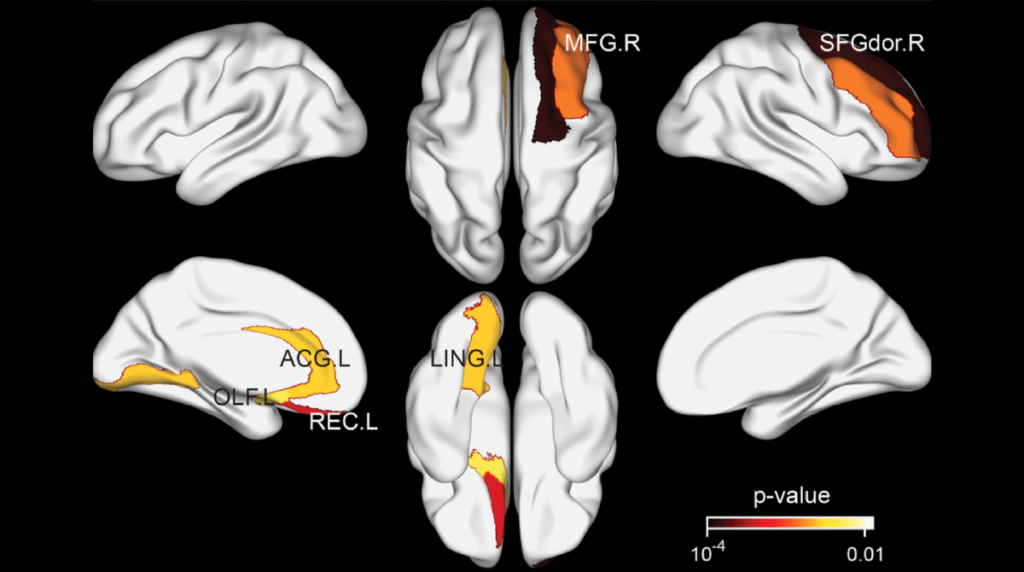
The researchers note that aging-related brain atrophy occurs in a predictable manner across the human lifespan. Therefore, brain atrophy is the measure of brain aging identified by MRI scans in this study. To translate the atrophy of brain structures into a biomarker of aging, the team leveraged machine-learning algorithms to generate a composite age-related morphological index called the Spatial Pattern of Atrophy for Recognition (SPARE) of Brain Age (SPARE-BA).

“The goal of the present study was to quantify the associations of epigenetic age acceleration and SPARE-BA acceleration with subsequent cognitive performance in a biracial cohort (~40% Black participants and ~60% White participants) of middle-aged adults with 5 to 15 years of follow up.”

The Results

Out of the four epigenetic aging markers examined, the researchers found that GrimAA was uniquely capable of closely predicting worse cognitive outcomes in this middle-aged CARDIA population. In the long term, biomarkers of epigenetic aging were more stable predictors of cognitive decline than the brain aging biomarker. However, changes in SPARE-BA and the SPARE-BA acceleration (SPARE-BAA) index showed stronger associations with cognition over time than any of the epigenetic aging markers. The researchers believe this is because the brain age/aging biomarkers may be more temporally dynamic in association with cognitive decline. When the researchers compared each biomarker’s association with cognition, they found that a combined model of GrimAA and SPARE-BAA demonstrated an improved ability to predict lower cognitive performance.

“GrimAA and SPARE-BAA were not correlated with one another, indicating that they capture distinct facets of biological aging.”

Conclusion

The researchers were forthcoming about limitations in this study. The epigenetic and brain imaging markers were mostly derived from different participants within the study, therefore, other unmeasured factors may have contributed to the study results. Baseline cognitive data was not recorded at younger ages and epigenetic markers were collected at different time points than cognitive and neuroimaging outcomes. These differences inhibited cross-sectional analysis of epigenetic and brain aging. In addition, predictions may be better validated with extended follow-up periods. Nonetheless, this research may have identified two profoundly useful indicators of cognitive decline that could be put to use as early as middle-age—a potential “tipping point” in the human lifespan; when interventions may still prevent irreversible cognitive impairment.

“With further validation, epigenetic and brain aging markers may help aid timely identification of individuals at risk for accelerated cognitive decline and promote the development of interventions to preserve optimal functioning across the lifespan.”

Click here to read the full research paper published by Aging (Aging-US).

AGING (AGING-US) VIDEOS: YouTube | LabTube | Aging-US.com

Aging (Aging-US) is an open-access journal that publishes research papers bi-monthly in all fields of aging research. These papers are available to read at no cost to readers on Aging-us.com. Open-access journals offer information that has the potential to benefit our societies from the inside out and may be shared with friends, neighbors, colleagues, and other researchers, far and wide.

For media inquiries, please contact media@impactjournals.com.

Trending With Impact: Can Job Stress Cause Epigenetic Aging?

The association between job-related stress and epigenetic aging was investigated using five epigenetic clocks and a Finnish cohort.

Job stress

The Trending With Impact series highlights Aging (Aging-US) publications that attract higher visibility among readers around the world online, in the news, and on social media—beyond normal readership levels. Look for future science news about the latest trending publications here, and at Aging-US.com.

Listen to an audio version of this article

In aging research, recent evidence has encouraged more focus on investigating socioeconomic status (SES) and its role in human health trajectories. Previous studies have used DNA methylation measures and epigenetic clocks to demonstrate a consistent association between low SES and epigenetic age acceleration (EAA). Moreover, researchers have identified a need to further investigate the relationship between SES characteristics and aging.  

“Little is known whether current occupational characteristics or job-related stress – crucial SES characteristics – are associated with EAA.”

Recently, researchers—from Imperial College LondonUniversity of SassariUniversity of Eastern FinlandKarolinska InstitutetUniversity of Oulu, and the Italian Institute for Genomic Medicine—conducted a research study in an effort to help elucidate potential mechanisms by which work characteristics and job stressors may be impacting health and accelerating aging. Their trending research paper was published by Aging (Aging-US) on February 2, 2022, and entitled, “Work-related stress and well-being in association with epigenetic age acceleration: A Northern Finland Birth Cohort 1966 Study.” 

The Study

The researchers in this study included 604 participants from the Northern Finland Birth Cohort 1966. Participants in this cohort were all born in the provinces of Oulu and Lapland, Finland, in 1966. DNA samples were collected and used to determine the relationship between biomarkers of aging, job stress and common environmental factors associated with age acceleration, including obesity, smoking, alcoholism, education status, and physical activity. The team used five different epigenetic clocks as biomarkers of aging: HorvathAA, HannumAA, PhenoAgeAA, GrimAgeAA, and DunedinPoAm.

“In this work, we assessed the association (and its magnitude) of five biomarkers of epigenetic age acceleration with work-related stress and well-being indicators (as well as other employment characteristics) in the Northern Finland Birth Cohort 1966, at 46 years old.”

Participants also filled out a clinical examination questionnaire, a modified Karasek’s Job Content Questionnaire (to assess job strain) and the Occupational Stress Questionnaire (to measure effort-reward imbalance). A number of descriptive statistics were collected from each participant, including body mass index (BMI); educational level; alcohol consumption; smoking habits; physical/leisure activity; job status (employed/unemployed); employer type (private or state/municipality); occupational group (white-collar or blue-collar); and job exposure. The researchers defined “job exposure” as job strain, effort-reward imbalance, overcommitment, occupational physical activity, work-favoring attitude, job security and work engagement, history, hours, and shift. 

The Results

After using linear regression models to analyze the adjusted and unadjusted pooled data (males and females together), the researchers found that job strain was not significantly associated with EAA using any of the epigenetic clocks. All five clocks associated smoking and obesity with accelerated aging (at varying significance). However, alcohol use (even heavy use) was not significantly associated with accelerated aging on any of the clocks. PhenoAgeAA associated job strain, active work and white-collar work (compared to blue-collar) with decreased aging. According to the Hannum and HorvathAA biomarkers of aging, people who worked more than 40 hours per week showed increased EAA.

“Once we stratified analyses by sex, a different pattern of association emerged, with women leading on the statistically significant results.”

Next, the researchers further stratified the results by sex. In men, high-intensity physical effort at work had a decreased aging effect. However, for women, high-intensity physical effort at work had an increased aging effect. The researchers point out that these clocks may have contradictory result due to the fact that women and men often present with diverse, sex-specific epigenetic patterns. While a direct correlation between job stress and epigenetic aging have yet to be proven, the degree of association between work characteristics and biomarkers of epigenetic aging in this study did vary by sex.

Conclusion

“This paper is one of the first attempts to address the working dimension of epigenetic age acceleration indicators, to the best of our knowledge.”

The Northern Finland Birth Cohort 1966 is a useful sample for studying a general population, and many confounders were removed in doing so. However, the researchers were forthcoming about some limitations that remained in this study. The unique characteristics of the cohort, as well as the questionnaires, may be responsible for the results seen in the study. The researchers suggest that additional studies be carried out in other societies and on different types of jobs to account for gender differences. 

“Our results suggest that women and men present different associations with different epigenetic distributions regarding work-related stress indicators.”

Click here to read the full research paper published by Aging (Aging-US).

AGING (AGING-US) VIDEOS: YouTube | LabTube | Aging-US.com

Aging (Aging-US) is an open-access journal that publishes research papers bi-monthly in all fields of aging research. These papers are available to read at no cost to readers on Aging-us.com. Open-access journals offer information that has the potential to benefit our societies from the inside out and may be shared with friends, neighbors, colleagues, and other researchers, far and wide.

For media inquiries, please contact media@impactjournals.com.

Trending With Impact: Radiation, Senescence and Senotherapeutics

Researchers examined the effects of thoracic radiation-induced senescent cells on tumor progression, and the role of senotherapeutics to mitigate these effects.

Radiation therapy, advanced medical linear accelerator in therapeutic oncology to treat cancer

The Trending With Impact series highlights Aging (Aging-US) publications that attract higher visibility among readers around the world online, in the news, and on social media—beyond normal readership levels. Look for future science news about the latest trending publications here, and at Aging-US.com.

Listen to an audio version of this article

Radiation therapy is a highly-efficacious inducer of cancer cell death. With this being said, radiation has also previously been shown to cause premature senescence in the lung parenchyma. Senescence in cancer cells was previously only thought of as a mechanism capable of suppressing tumor cell proliferation by halting the cell cycle. However, a growing body of evidence shows that senescent cells may play a pro-tumorigenic role in cancer.

In the tumor microenvironment, the accumulation of senescent cells can become tumorigenic due to a lack of normal tissue stem cells and due to the expression of the senescence-associated secretory phenotype (SASP). SASP expression is when senescent cells secrete high levels of inflammatory cytokines, immune modulators, growth factors, and proteases. In addition to reinforcing senescence, SASP can create a biological environment that is immuno-suppressed and tumor-permissive. Radiation-induced senescence has previously been shown to have negative impacts on cancer patients.

“Cells that have undergone premature senescence due to stress, such as irradiation, are resistant to apoptotic cell death and effectively escape immune surveillance, resulting in their accumulation in tissue over time.”

Recently, researchers from the National Cancer Institute investigated the irradiated lung and the impact of radiation-induced senescent parenchymal cells on tumor growth. They also explored three senotherapeutics, rapamycin, INK-128 and ABT-737, for their potential to mitigate radiation-induced senescence. On February 12, 2022, the team’s priority research paper was published on the cover of Aging (Aging-US) Volume 14, Issue 3, and entitled, “Senescence-associated tumor growth is promoted by 12-Lipoxygenase.”

The Study

In this study, researchers intravenously injected melanoma cells into murine models two, four and eight weeks after daily fractions of thoracic irradiation exposure. There was also a control arm of unirradiated murine models. Tumor development was monitored by the number and size of the nodules in lung tissues. The number of cells exhibiting senescent activity was also recorded after two, four and eight weeks of thoracic irradiation. Their data demonstrated a correlation between the time points when tumors developed in the irradiated lungs and a marked accumulation of senescent cells.

“As previously described, in irradiated lungs, senescent cells increased significantly 4 and 8 weeks after IR compared to age matched unirradiated controls (Figure 1A).”

A characteristic of oncogene- and stress-induced senescence is the activation of mTOR signaling. Given this connection, the researchers conducted parallel studies evaluating senostatic agents capable of targeting the mTOR pathway, rapamycin and INK-128, and a senolytic agent to selectively eliminate senescent cells, ABT-737.  The data showed that rapamycin and INK-128 significantly reduced the number of tumor nodules in the lungs of irradiated mice compared to the controls. ABT-737 demonstrated reduced pulmonary senescence in irradiated mice.

The researchers also studied 12-Lipoxygensae (12-LOX), an enzyme that metabolizes a certain SASP molecule previously implicated in pulmonary senescence: 12(S)-HETE. 12-LOX is a known contributor to radiation-induced senescence and lung injury. The team specifically focused on the role of 12-LOX in pulmonary senescence and its impact on radiation-enhanced tumor growth. They found that inhibiting 12-LOX activity reduced radiation-induced lung senescence and mitigated radiation-enhanced tumor growth.

“Finally, we link senescence associated 12-LOX activity and production of 12(S)-HETE to the observed enhanced tumor growth after irradiation.”

Conclusion

In sum, the researchers found that radiation therapy can induce senescence in the lung parenchyma and also enhance tumor growth. The contribution of senescence in tumor progression was emphasized by the protection delivered by the mTOR-targeted senostatic and senolytic agents. This important discovery could lead to new therapies for cancer patients who are undergoing radiation therapy.

“Together, this study demonstrates the critical role of senescence in mediating radiation-enhanced tumor growth and identifies Alox12 as an important player in this phenomenon. Treatment with a senostatic agent, INK-128, identified in this study, or with agents like rapamycin and ABT-737 suggested their potential therapeutic use in alleviating radiation associated tumor growth.”

Click here to read the full priority research paper published by Aging (Aging-US).

AGING (AGING-US) VIDEOS: YouTube | LabTube | Aging-US.com

Aging (Aging-US) is an open-access journal that publishes research papers bi-monthly in all fields of aging research. These papers are available to read at no cost to readers on Aging-us.com. Open-access journals offer information that has the potential to benefit our societies from the inside out and may be shared with friends, neighbors, colleagues, and other researchers, far and wide.

For media inquiries, please contact media@impactjournals.com.

Trending With Impact: ARDD21 Meeting Report Highlights

Read a brief summary of a meeting report from the 8th Annual Aging Research and Drug Discovery (ARDD21) meeting. 

ARDD21

The Trending With Impact series highlights Aging (Aging-US) publications that attract higher visibility among readers around the world online, in the news, and on social media—beyond normal readership levels. Look for future science news about the latest trending publications here, and at Aging-US.com.

Listen to an audio version of this article

The 8th Annual Aging Research and Drug Discovery (ARDD21) meeting was held in Copenhagen, Denmark, from August 30 to September 3, 2021. This meeting was attended by over 130 people on-site, with an additional 1800 people engaged online. The focus of this meeting was the current landscape of aging research and various ways it can be applied to drug discovery. Topics included: age-dependent control of cellular maintenance processes, longevity pathways, artificial intelligence-based drug screening, cellular stress and aging, the benefits of dietary restriction, stem cell rejuvenation, senolytics as an aging therapeutic, diverse models of aging, aging clocks and biomarkers of aging, new ideas in preclinical and clinical aging research, the longevity industry landscape, and a Longevity Medicine Workshop.

In total, there were 75 presentations given at ARDD21 by prominent and dedicated aging researchers. The meeting was thoroughly summarized in a paper published in Aging (Aging-US) Volume 14, Issue 2, entitled, “Meeting Report: Aging Research and Drug Discovery.”

ARDD21 Meeting Report Highlights

One of the keynote presentations was given by Nir Barzilai from the Albert Einstein College of Medicine. He discussed his work on aging and how it can be applied to drug discovery. One interesting finding that he discussed was that many drugs currently used to treat chronic diseases, such as diabetes and heart disease, also have the potential to treat aging. This is due to the fact that many diseases are symptoms of aging, and thus, treating the underlying cause (aging) can in turn treat the symptoms.

Another keynote presentation was given by James Kirkland from the Mayo Clinic. He discussed his work on developing therapies to target senescent cells. Senescent cells can accumulate with age, and their presence has been linked with a variety of age-related conditions such as arthritis, cancer and heart disease. Kirkland’s team has developed a number of potential therapies to eliminate or reduce the number of senescent cells in the body, and he is currently testing them in clinical trials.

Professor Dame Linda Partridge from Max Planck Institute for Biology of Ageing presented on aging and the importance of intestinal homeostasis. Her studies involved rapamycin treatment to act on the longevity pathway mTOR, which revealed that short term and early treatment with rapamycin extends lifespan in D. melanogaster as much as chronic rapamycin treatment. Yu-Xuan Lu, another researcher from the Max Planck Institute for Biology of Ageing, demonstrated the existence of an unconventional intestine sex-specific TORC1-histone axis which uncovers a new aspect of improved longevity with rapamycin.

Brian Kennedy from the Buck Institute for Research on AgingNational University of Singapore and National University Health System showed how Alzheimer’s disease can be used as a model of neuronal aging. Presenting their new WormBot, Matt Kaeberlein from the University of Washington described a “set it and forget it” method of large-scale intervention testing in roundworms (C. elegans). 

“He stressed the importance of broad and unbiased screening of intervention beyond known pathways and in different combinations [56].”

Aging (Aging-US) Editorial Board member Alexey Moskalev from the Russian Academy of Sciences presented on the disruption of hydrogen sulfide homeostasis and its association with aging, and therefore, its potential as a gero-therapeutic target. David Sinclair from Harvard Medical School (also on the Aging Editorial Board) discussed aging-driven epigenetic and gene expression changes in the central nervous system. He showed that this can be safely reversed to restore vision by inducible adeno-associated viruses expressing polycistronic Oct4, Sox2 and Kif4, and that the effect is dependent on DNA demethylation. Finally, a Longevity Medicine Workshop was held with a panel of experts aimed to inspire young students to engage in longevity research. This panel included Aging Editorial Board members Alex Zhavoronkov, Alexey Moskalev and Mikhail Blagosklonny (Editor-In-Chief).

Conclusion

Overall, the ARDD21 meeting was a fruitful exhibition of experts from all areas of aging research that came together to share their latest findings in the field. The highlights in this blog pale in comparison to the thoughtful details included in the original meeting report. 

Click here to read the full meeting report published by Aging (Aging-US).

AGING (AGING-US) VIDEOS: YouTube | LabTube | Aging-US.com

Aging (Aging-US) is an open-access journal that publishes research papers bi-monthly in all fields of aging research. These papers are available to read at no cost to readers on Aging-us.com. Open-access journals offer information that has the potential to benefit our societies from the inside out and may be shared with friends, neighbors, colleagues, and other researchers, far and wide.

For media inquiries, please contact media@impactjournals.com.

Trending With Impact: Therapeutic Strategy Improves Cell Senescence

In the cover paper of Aging (Aging-US) Volume 14, Issue 2, researchers discovered a potential therapeutic strategy to target senescent cells and combat aging and age-related diseases.

The Trending With Impact series highlights Aging (Aging-US) publications that attract higher visibility among readers around the world online, in the news, and on social media—beyond normal readership levels. Look for future science news about the latest trending publications here, and at Aging-US.com.

Listen to an audio version of this article

Cellular senescence appears to be a phenomenon fundamentally ingrained within the aging process and linked to age-related diseases. Characterized broadly by permanent cessation of the cell cycle, cellular senescence may not be as permanent as once thought. 

Researchers from Incheon National University and Korea University conducted a new study exploring analogs of oxazoloquinoline and their potential to alleviate cellular senescence. Their trending research paper was published as the cover of Aging (Aging-US) Volume 14, Issue 2, and entitled, “Targeting regulation of ATP synthase 5 alpha/beta dimerization alleviates senescence.”

THE STUDY

Adenosine triphosphate (ATP) is an energy-carrying molecule found in all living cells. In order to meet the energy demands of the cell, the primary function of the mitochondria is to produce ATP. The maintenance of mitochondrial metabolism is inseparably linked with the regulation of senescence. Therefore, dysfunctional mitochondria has been considered as both a target and the cause of senescence. In addition to a marked decrease in ATP production, senescent cells also increase the expression of inflammatory cytokines, including interleukin 33, or IL-33. The researchers believe that reducing IL-33 may be a possible intervention to reduce senescence in aging patients and age-related diseases.

“In this study, using in-house compound library containing 20 oxazoloquinoline analogs designed to IL-33 inhibitors [9], we aimed to identify compounds capable of ameliorating senescence.”

The researchers investigated 20 oxazoloquinoline analogs using in vitro assays of senescent human diploid fibroblasts and embryonic kidney cells. Efficacy of the candidate compounds was determined using a screening strategy to measure their capacity to increase cell number. Cell numbers were measured between zero and 20 days after compound exposure. The researchers also measured indicators including mitochondrial membrane potential, reactive oxygen species (ROS) levels and p21 expression. They found that the analog KB1541 led to the maximum cell number increase, the recovery of mitochondrial function and the alleviation of cellular senescence. The researchers suggest that KB1541 could be a promising therapeutic agent for use in aging-related diseases.

“The increase in mitochondrial cristae length by KB1541 could be explained by previous findings showing that the increase in ATP generation exerted beneficial effects in mitochondrial function including increases in calcium buffering capacity and decrease in overall ROS production [48].”

CONCLUSIONS

“Taken together, our study provides evidence that the fine-tuning of ATP synthase 5 alpha/beta dimerization by KB1541 can induce mitochondrial functional recovery, concomitant recovery of senescent phenotypes, rendering the use of KB1541 as a potentially advantageous therapeutic strategy in aging and age-related diseases.”

The authors acknowledged that further studies are needed to clarify the exact relationship between IL-33 and mitochondrial energy metabolism. Further studies are also needed to investigate whether other IL-33 inhibitors can modulate senescence by the mechanisms found in the study. This research provides valuable insight into the potential of oxazoloquinoline analogs as novel therapeutic agents for aging and age-related diseases. With further exploration, their findings could lead to new therapeutic strategies to combat aging.

“The role of IL-33 in senescence is not clearly elucidated, therefore discovery of a novel interacting partner will provide clues toward revealing its function.”

Click here to read the full research paper published by Aging (Aging-US).

AGING (AGING-US) VIDEOS: YouTube | LabTube | Aging-US.com

Aging (Aging-US) is an open-access journal that publishes research papers bi-monthly in all fields of aging research. These papers are available to read at no cost to readers on Aging-us.com. Open-access journals offer information that has the potential to benefit our societies from the inside out and may be shared with friends, neighbors, colleagues, and other researchers, far and wide.

For media inquiries, please contact media@impactjournals.com.

How Habitual Tea Drinking Impacts Brain Structure

In 2019, researchers conducted the first study to explore the effects of habitual tea drinking on system-level brain networks.

Figure 3. Brain regions exhibiting significant differences in structural nodal efficiency between the tea drinking group and the non-tea drinking group at the significance level of 0.01 (uncorrected) statistical evaluated by a permutation test.
Listen to an audio version of this article

After water, tea is the most popular beverage in the world. While many people enjoy tea for the flavor, aroma and caffeine boost, research suggests that there may be another reason to regularly drink this beverage: its effects on the brain. In 2019, researchers from Wuyi UniversityUniversity of EssexUniversity of Cambridge, and the National University of Singapore conducted the first study exploring the effects of tea on system-level brain networks. Their paper was published in Aging (Aging-US) Volume 11, Issue 11, and entitled, “Habitual tea drinking modulates brain efficiency: evidence from brain connectivity evaluation.”

“In this study, we comprehensively explored brain connectivity with both global and regional metrics derived from structural and functional imaging to unveil putative differential connectivity organizations between tea drinking group and non-tea drinking group.”

The Study

The subjects enrolled in this study were older adults (mean ≈ 70 years old) from residential communities in Singapore, without conditions or terminal illnesses (see Materials and Methods). Researchers initially recruited 93 participants, however, only 36 total participants (male = 6; female = 30) remained after adjusting for the strict study inclusion criteria. Researchers classified the remaining participants as “non-tea drinkers” or “tea drinkers” using complex composite test scores. The composite score included self-reports of multiple decades of weekly green tea, oolong tea, black tea, and coffee intake (see Materials and Methods). After screening, 15 participants were assigned to the tea-drinking group and 21 were assigned to the non-tea drinking group. (Coffee intake did not differ significantly between the two groups.)

Next, structural brain connectivity was compared between tea drinkers and non-tea drinkers. All 36 participants underwent MRI brain scans and both functional and structural networks were investigated from global and regional perspectives. The researchers found that participants in the tea-drinking group had more efficient structural organization. However, tea did not seem to have a significantly beneficial effect on global functional organization. As a result of tea drinking, hemispheric asymmetry in the structural connectivity network was observed, although it was not observed in the functional connectivity network. 

“In addition, functional connectivity strength within the default mode network (DMN) was greater for the tea-drinking group, and coexistence of increasing and decreasing connective strengths was observed in the structural connectivity of the DMN.”

Conclusion

The researchers found that tea drinkers had more efficient brain structure organization than non-tea drinkers. Studies have previously demonstrated that tea drinkers are less likely to develop dementia, and tea consumption has also been linked with better cognitive performance. The researchers note that these effects are due to tea’s contents of caffeine, L-theanine and polyphenols (catechins). Polyphenols are compounds found in plants, including tea leaves, and may help protect against oxidative damage. Previous studies have shown that tea polyphenols can cross the blood-brain barrier and may help improve brain function. 

While this study’s findings suggest that habitual tea drinking leads to better brain connectivity and efficiency in old age, the researchers were forthcoming about the limitations of their study. The sample size was limited and other substances, behaviors, habits, and environmental factors may have impacted the outcome of the study. 

“Our study offers the first evidence of the positive contribution of tea drinking to brain structure and suggests a protective effect on age-related decline in brain organisation.”

Click here to read the full research paper published by Aging (Aging-US).

AGING (AGING-US) VIDEOS: YouTube | LabTube | Aging-US.com

Aging (Aging-US) is an open-access journal that publishes research papers monthly in all fields of aging research and other topics. These papers are available to read at no cost to readers on Aging-us.com. Open-access journals offer information that has the potential to benefit our societies from the inside out and may be shared with friends, neighbors, colleagues, and other researchers, far and wide.

For media inquiries, please contact media@impactjournals.com.

Investigating a Biomarker of Age-Related Macular Degeneration

Age-related macular degeneration
Ocular drusen in age-related macular degeneration (AMD).

Over 15 million people in the United States are currently struggling with age-related macular degeneration (AMD). Among aging populations around the world, AMD is the leading cause of irreversible vision loss. A main target of AMD is the pigmented layer of the retina, or the retinal pigment epithelium (RPE). The subretinal space of the eye encompasses the area under the retina between RPE cells and photoreceptors. Yellow deposits of lipids and proteins, called drusen, are located in the subretinal space and the hallmark sign of AMD. In 2005, researchers found that drusen deposits also contain an immune protein called the C-reactive protein (CRP).

Circulating CRP is one of the main clinical biomarkers of inflammation and infection, since CRP levels rise and fall with inflammation. This immune-response protein can exert pro-inflammatory properties by disassociating from its pentameric form (pCRP) into smaller monomeric CRP (mCRP) subunits. These mCRPs are small enough to cross the ocular blood–retinal barrier (oBRB) and appear in ocular drusen.

“mCRP has been identified in ocular drusen and other subepithelial deposits [2425], as well as in the choroid, and we have shown that mCRP, but not pCRP, contributes to oBRB disruption in vitro [26].”

Researchers—from Hospital Clínic de BarcelonaManchester Metropolitan UniversityHospital de la Santa Creu i Sant PauInstitute Salud Carlos IIIUniversity of BristolMoorfields Eye Hospital, and University College London Institute of Ophthalmology—conducted a study aimed at understanding the mCRP’s contribution to the pathophysiology of AMD. Their paper was published by Aging (Aging-US) in 2020, and entitled, “Activation of C-reactive protein proinflammatory phenotype in the blood retinal barrier in vitro: implications for age-related macular degeneration”.

The Study

“If mCRP pro-inflammatory capacity is unrestrained in AMD and particularly in high risk patients, then we need to determine how mCRP is generated or accumulates in the subretinal space as there is no CRP transcription in the retinal tissue [3031].”

In order to investigate how mCRP is generated and/or how it accumulates within the subretinal space, the researchers used a Transwell model to first determine whether circulating CRP could reach the subretinal space. The Transwell model included monolayers of primary porcine choroidal endothelial cells (CECs) grown on porous filters with their apical and basolateral surfaces exposed to separate chambers. They found that CRP isoforms were able to cross the CEC monolayer on the apical side of the RPE. Next, they used the Transwell model to evaluate whether CRP isoforms could also reach the subretinal space and cross RPE ARPE-19 cells. They found that mCRP was able to diffuse into both the subretinal space and cross into the RPE. pCRP was not found in the opposite chambers, even after 24 and 48 hours post-exposure. However, when the researchers tested younger and healthier primary porcine RPE cells instead of the ARPE-19 cells, neither pCRP nor mCRP diffused into opposite chambers.

Next, the researchers studied whether pCRP could dissociate into mCRP within the RPE. They found that induced inflammation triggered pCRP dissociation into mCRP in both the ARPE-19 and primary porcine RPE cells. The team also found that barrier disruption induced by mCRP was dependent on its topological localization.

Conclusion

“In summary, our findings further support mCRP direct contribution to progression of AMD, at least at the RPE level. The topological experiments elicit that mCRP is proinflammatory when present on the apical side of the RPE. However, mCRP is likely to only reach the apical side of the RPE in compromised RPE health and where barrier functions are compromised.”

The researchers were forthcoming about limitations in their study. Nonetheless, this study suggests that a plausible mechanism by which mCRP may contribute to RPE dysfunction and AMD progression is, when pCRP reaches the oBRB, it diffuses past the oBRB by dissociating into mCRP. It is also possible that mCRP may be derived from the dissociation of pCRP on the surface of damaged RPE. The proinflammatory microenvironment may be amplified and the barrier disruption may be enhanced when mCRP reaches the apical side of an already aged or damaged RPE.

“With respect to previous findings, this pathologic mechanism will be more prevalent in patients carrying the FH risk polymorphism for AMD, where mCRP proinflammatory effects remain unrestrained [28].”

Click here to read the full research paper published by Aging (Aging-US).

AGING (AGING-US) VIDEOS: YouTube | LabTube | Aging-US.com

Aging (Aging-US) is an open-access journal that publishes research papers bi-monthly in all fields of aging research and other topics. These papers are available to read at no cost to readers on Aging-us.com. Open-access journals offer information that has the potential to benefit our societies from the inside out and may be shared with friends, neighbors, colleagues, and other researchers, far and wide.

For media inquiries, please contact media@impactjournals.com.

Trending With Impact: Hair Follicles May Replace Traditional Biopsies

A new device has been developed by researchers to efficiently and painlessly collect hair follicle tissue samples from laboratory mammals, and even humans.

Figure 6. Markers of senescence analysis in hair follicular cells.

The Trending With Impact series highlights Aging (Aging-US) publications that attract higher visibility among readers around the world online, in the news, and on social media—beyond normal readership levels. Look for future science news about the latest trending publications here, and at Aging-US.com.

Laboratory mammals have impacted human-kind far beyond enhancing scientific knowledge in behavioral and environmental research. These animals have greatly contributed to human healthspan and lifespan in countless ways; from validating life-saving cancer therapies to accelerating the future of human anti-aging and longevity interventions. With respect for these salubrious animals, ethical standards (per country) require that researchers handle laboratory mammals with care, and that pain and stress are minimized. Blood and skin tissue samples (biopsies) collected from animals should be replaced whenever possible. For the researchers, this twofold invasive procedure for the animals is also time- and resource-limiting—presenting a bottleneck in the biomedical research process.

“However, we present here a simple method for obtaining biological material in the form of follicular cells from laboratory mice with sufficient quantities and quality for multiple analyses using standard modern molecular biology methods.”

In an effort to efficiently and humanely solve this ethics/logistics problem, researchers—from Palacky UniversityUniversity Hospital OlomoucDanish Cancer Society Research Center, and Karolinska Institute—developed a novel, non-invasive device that can be used to collect tissue samples from hair follicles. They tested the applications of this device and authored a research paper of the study. In December of 2021, their paper was published on the cover of Aging (Aging-US) Volume 13, Issue 23, and entitled, “An efficient, non-invasive approach for in-vivo sampling of hair follicles: design and applications in monitoring DNA damage and aging.”

“As millions of laboratory mice are routinely genotyped globally every year this approach represents a major ethical and logistic breakthrough.”

The Follicular Cells’ Collector

As opposed to traditional biopsies, hair follicle collection is a humane, easy, non-invasive, and painless method of DNA and tissue sample collection. Each hair follicle contains approximately 50 cells—of various cell types. 

“This micro-organ structure also has other advantages in biomarker studies, including suitability for investigations of circadian rhythms [57], and the presence of numerous cell types in a small area, which can be easily distinguished, such as keratinocytes, melanocytes, or perifollicular macrophages and mast cells [810].”

Previously, the limitations of using hair follicles as DNA and tissue samples stemmed from ineffective technology. Many devices involved ordinary tweezers and forceps with high risks for cross-contamination. The researchers termed their novel tissue sample collection device the “follicular cells’ collector.” The follicular cells’ collector is designed with dual pipettes and utilizes a precision vacuum method of hair follicle extraction. The device can be used to comfortably collect DNA and tissue samples from laboratory mammals, and even from humans. 

“Although hair samples have been previously used for that purpose [2931], our sample collection approach may motivate researchers to use them more routinely and widely.”

The Study

To validate that these hair follicle samples contain the required genetic information necessary in most studies, researchers compared murine genotyping results of 151 tail biopsies and 151 hair samples. In order to determine the ability of these samples to detect changes in expression patterns induced by external factors, the team also observed the DNA damage response in hair follicle cells after gamma irradiation and after the topical application of chemical clastogens. Further exploring its potential application in aging research, researchers assayed expression patterns of selected markers of biological age and senescence in murine hair follicular cells. The researchers conducted many other tests and experiments using murine hair follicular cells in this study.

“The speed by which the samples can be collected and processed (e.g. by fixation) is among the biggest advantages of our solution as it can be performed within seconds. This fact limits any potential underlying cellular responses and additional DDR [DNA damage response] caused by cofounding stressing factors related to the withdrawal process [2].”

Conclusion

The researchers found that the follicular cells’ collector method of obtaining mouse hair follicular cells can be successfully used for genotyping, quantitative polymerase chain reaction testing and quantitative immunofluorescence. They also demonstrated that this method can successfully monitor quality and expression level changes of selected proteins—induced by external factors and during natural or experimentally induced aging. 

“Our results highlight the value of hair follicles as biological material for convenient in vivo sampling and processing in both translational research and routine applications, with a broad range of ethical and logistic advantages over currently used biopsy-based approaches.”

Click here to read the full research paper published by Aging (Aging-US).

AGING (AGING-US) VIDEOS: YouTube | LabTube | Aging-US.com

Aging (Aging-US) is an open-access journal that publishes research papers bi-monthly in all fields of aging research and other topics. These papers are available to read at no cost to readers on Aging-us.com. Open-access journals offer information that has the potential to benefit our societies from the inside out and may be shared with friends, neighbors, colleagues, and other researchers, far and wide.

For media inquiries, please contact media@impactjournals.com.

Trending With Impact: Aging Reduced by 8 Years With Rejuvant®

A recent study revealed that participants experienced an average 8 year reduction in biological aging after taking Rejuvant® for approximately 7 months.

Anti-aging hourglass

The Trending With Impact series highlights Aging (Aging-US) publications that attract higher visibility among readers around the world online, in the news, and on social media—beyond normal readership levels. Look for future science news about the latest trending publications here, and at Aging-US.com.

Listen to an audio version of this article

Waiting until the end of a subject’s lifespan is quite a leaden method of validating the efficacy of a longevity-based intervention. This method could take researchers generations upon generations to eventually validate an effective intervention—or—this method might not ever yield results seen by the general public. However, researchers may have devised an innovative way to solve this problem.

“If we hope to control the aging process, we need to learn how to measure the rate of aging in shorter time periods.”

Many researchers believe that measuring the rate of human aging can be done faster by using DNA methylation-based aging clocks. Methylation-based clocks are capable of determining human biological aging with impressive accuracy. Hypermethylated and demethylated regions of DNA (CpG islands near specific aging-associated genes) play a key role in turning certain genes on and off throughout the aging process. Therefore, methylation is a biomarker of aging. While there is a short list of currently available biological aging clocks for researchers to use in studies of anti-aging therapies, the TruAge DNA methylation test is preferable in some cases, due to its accessibility, use of simple saliva samples and cost effectiveness.

“For the first time, these biomarkers of aging give scientists the opportunity to study the effects of anti-aging compounds in real-time and directly in humans.”

In a new study, researchers from TruMe LabsNational University of Singapore and Ponce de Leon Health used the TruAge DNA methylation test to validate Rejuvant®—a patent-pending anti-aging dietary supplement. The trial study yielded unprecedented results and the research paper authored by the team was published as the cover of Aging (Aging-US) Volume 13, Issue 22, entitled: “Rejuvant®, a potential life-extending compound formulation with alpha-ketoglutarate and vitamins, conferred an average 8 year reduction in biological aging, after an average of 7 months of use, in the TruAge DNA methylation test”.

The Study

Developed at Ponce de Leon Health, Rejuvant® is composed primarily of a compound called Alpha-Ketoglutarate (AKG). This molecule is naturally produced in humans and functions as a signaling molecule, an energy donor, a precursor to amino acid biosynthesis, and a regulator of epigenetic processes. In humans and other animals, AKG levels gradually decrease with age. Other components of Rejuvant® are calcium and, for males and females, Vitamin A and Vitamin D, respectively. 

“The goal of the study was to determine the effect of Rejuvant® supplementation on human biological aging by measuring DNA methylation.”

Researchers enrolled 42 healthy participants who were on average 64 years of age (43 to 72). Before taking Rejuvant®, all 42 participants completed a survey and their baseline biological age was measured using the TruMe age prediction model. The survey was a self-reported questionnaire including information about diet, alcohol intake, previous consumption of Rejuvant®, health, height and weight, sleep duration, smoking status, exercise frequency, physical activity level, meal frequency, snacking frequency, number of additional dietary supplements consumed and frequency, hair status, education, healthy lifestyle mindset, and trust in dietary supplements. 

Participants (majority male; 28) took two tablets of Rejuvant® daily, for a duration of four to 10 months. Biological age was measured from saliva samples again after taking Rejuvant® for four to 10 months. At the end of the trial, participants completed the same survey. The researchers compared the baseline surveys with the final surveys to check for other confounders contributing to the results in the study. They also used the surveys to select a sub-group of 13 participants who reported no change in diet type, drinking frequency, additional dietary supplements intake, sleep duration, and exercise frequency. They compared this sup-group with the rest of the cohort. They also compared results between males and females, older and younger participants, and participants with higher biological age relative to their chronological age (aging more quickly).

Results and Conclusion

Researchers examined associations between the epigenetic clock, health status, physical fitness, and the effects of Rejuvant® on human biological aging. The researchers were forthcoming about limitations in this study. A control arm was not used, the cohort was relatively small, only one biological aging clock was used, and researchers did not collect other kinds of data relevant to aging. However, the study results showed that Rejuvant® conferred an average eight year reduction in biological aging after approximately seven months of use. The 13 participants in the sub-group saw anti-aging benefits slightly less than the rest of the cohort. Rejuvant® was more effective in chronologically older participants and in participants that were aging more quickly (with a higher biological age relative to their chronological age).

“Future randomized clinical trials will be required to confirm the findings presented here. Nevertheless, the results in this manuscript suggest that Rejuvant® may have significant effects on biological age as measured by DNA methylation of saliva samples.”

Click here to read the full priority research paper published by Aging (Aging-US).

WATCH: AGING VIDEOS ON LABTUBE

Aging (Aging-US) is an open-access journal that publishes research papers monthly in all fields of aging research and other topics. These papers are available to read at no cost to readers on Aging-us.com. Open-access journals offer information that has the potential to benefit our societies from the inside out and may be shared with friends, neighbors, colleagues, and other researchers, far and wide.

For media inquiries, please contact media@impactjournals.com.

Trending With Impact: Worms Reveal Early Event in Neurodegeneration

Researchers examined roundworms to determine the role of mitochondrial dysfunction in progressive neurodegenerative disorders, such as Alzheimer’s disease.

From Figure 2. Altered mitochondrial morphology and activity in tauwt-expressing larvae. (truncated)

The Trending With Impact series highlights Aging (Aging-US) publications that attract higher visibility among readers around the world online, in the news, and on social media—beyond normal readership levels. Look for future science news about the latest trending publications here, and at Aging-US.com.

Listen to an audio version of this article

Many aging-associated neurodegenerative disorders, including Alzheimer’s disease, involve the aggregation of abnormal tau in nerve cells (neurons). Normally, tau proteins function to stabilize microtubules in the brain. Tauopathy occurs when tau proteins become misfolded and misshapen (which turns tau into toxic tau). They then continue to proliferate and bind to each other, forming tau oligomers. These tau oligomers are more toxic and have a greater potential to spread tau pathology. Before the tau pathology snowballs into neurodegenerative disorders, the events that lead up to abnormal tau have remained elusive to researchers. 

“While the association between tau levels and energy metabolism is established, it is not clear whether mitochondrial dysfunction is an early pathological feature of high levels of tau or a consequence of its excessive formation of protein aggregates.”

Previous studies have demonstrated an association between tau levels and mitochondrial metabolism, however, determining which one proceeds the other has yet to be fully illuminated. Shedding light on this subject, researchers—from the University of CopenhagenNational and Kapodistrian University of Athens and the National Institutes of Health’s National Institute on Aging—used a Caenorhabditis elegans (C. elegans; roundworm/nematode) model of tau to examine mitochondrial changes over time. Their paper was chosen as the cover of Aging (Aging-US) Volume 13, Issue 21, published in November of 2021 and entitled, “Alteration of mitochondrial homeostasis is an early event in a C. elegans model of human tauopathy”.  

The Study

“Here, we utilized transgenic nematodes expressing the full length of wild type tau in neuronal cells and monitored mitochondrial morphology alterations over time.”

To investigate the impact of tau on mitochondrial activity, neuronal function and organismal physiology, the researchers selected and cultured an already characterized nematode strain that expresses the full length of wild type human tau protein. They compared wild type nematodes with tau-expressing nematodes (at various ages) over time using a thrashing assay, mitochondrial imaging, worm tracking software, and western blot analysis. Calcium deregulation was also examined to determine whether or not it is implicated in the impairment of mitochondrial activity in the tau-expressing nematodes. They found that chelating calcium led to restored mitochondrial activity and suggested a link between mitochondrial damage, calcium homeostasis and neuronal impairment in this nematode model.

Figure 2. Altered mitochondrial morphology and activity in tauwt-expressing larvae.

Conclusion

“Our findings suggest that defective mitochondrial function is an early pathogenic event of tauopathies, taking place before tau aggregation and undermining neuronal homeostasis and organismal fitness.”

The researchers were forthcoming about limitations in their study, given the differences between human and nematode biology and pathology. Nevertheless, they found evidence that, in this nematode tauopathy model, neurotoxicity depends on protein alterations and mitochondrial dysfunction. Mitochondrial dysfunction takes place before high levels of tau are detected. Tau mutations may also modulate calcium homeostasis by influencing the main cellular storage sites—the endoplasmic reticulum and mitochondria.

“Investigating the tight interplay between tau oligomers and energy metabolism will enlighten new avenues for therapeutic strategies to slow or halt the progression of dementia-related diseases such as AD [Alzheimer’s disease].”

Click here to read the full priority research paper published by Aging (Aging-US).

WATCH: AGING VIDEOS ON LABTUBE

Aging (Aging-US) is an open-access journal that publishes research papers monthly in all fields of aging research and other topics. These papers are available to read at no cost to readers on Aging-us.com. Open-access journals offer information that has the potential to benefit our societies from the inside out and may be shared with friends, neighbors, colleagues, and other researchers, far and wide.

For media inquiries, please contact media@impactjournals.com.

  • Follow Us