Dry Eyes? It May be Immune Infiltration in Aging Lacrimal Glands

In a new editorial, researchers from Baylor College of Medicine artfully discuss the immune system’s role in dry eye disease. 

The lacrimal gland, found in the upper outer part of the eye’s hollow area, is an important gland that makes tears to protect the eye from infections. It’s split into two parts: one near the inside of the eyelid that can be seen when the eyelid is flipped, and another part with ducts lower in the eye that connects to its counterpart. In their fully functioning status, these ducts release fluid onto the surface of the eye. As humans age (especially women), the lacrimal gland gradually becomes infiltrated by aberrant immune cells and can ultimately lead to an uncomfortable condition known as dry eye disease.

“Burning and redness in the eyes, grittiness and blurry vision make life miserable and currently, eye drops with a variety of lubricant components and in the most severe cases, immunosuppressors, are the only therapies approved for this disease.”

In a well-written new editorial paper, researchers Claudia M. Trujillo-Vargas and Cintia S. de Paiva from the Department of Ophthalmology at Baylor College of Medicine artfully discuss their recent studies which shed light on the immune system’s role in dry eye disease. On August 11, 2023, their editorial was published in Aging’s Volume 15, Issue 15, entitled, “Our search of immune invaders in the aged lacrimal gland.”

Editorial Summary

The authors write that their research group has been dedicated to investigating the changes that occur in the lacrimal gland due to aging and focus on immunopathological alterations. Due to limited human samples, their studies have centered on understanding the infiltration of lymphocytes, specifically B and T cells, in aged mice’s lacrimal glands. This infiltration has been linked to increased dysfunction of the ocular surface. 

“In the search of mechanisms that can counteract the effects of the overwhelming immune infiltration, we started characterizing one of the main players of immune tolerance, the thymic-derived T regulatory cells (Tregs).”

The researchers and their team have a particular interest in thymic-derived T regulatory cells (Tregs), which play a key role in immune tolerance. Paradoxically, in the aged glands, these Tregs, while exhibiting markers for their suppressive function, display heightened differentiation, infiltrate the tissue, produce inflammatory cytokines, and demonstrate impaired suppressive capabilities. When transferred to immunodeficient recipients, these dysfunctional Tregs replicate lacrimal gland pathology. 

Aged lacrimal glands contain highly differentiated CD4+ T cells of the Th1 and Th17 phenotypes, which exhibit exhaustion and immunopathological features. This environment hampers Tregs’ ability to suppress immune responses. There’s also an increase in naïve CD4+ T cells and IgD+ B cells, suggesting a unique environment for the recruitment of inexperienced immune cells in the gland.

Ectopic lymphoid structures, resembling those found in aged tissues, are observed in the lacrimal gland, potentially contributing to immune dysregulation. Despite the concept of immune cells being unwelcome invaders, the lacrimal gland relies on immune cell influx for surveillance purposes, as it is highly vascularized. Nonetheless, with age, immune cell infiltration intensifies, accompanied by fibrosis, duct issues and gland atrophy. Interestingly, antigen-presenting cells diminish, adding to the peculiar immune environment.

In their running analogy to the movie “Men in Black,” the researchers explain that they are seeking effective therapies, akin to the “noisy crickets,” to combat this pathological immune infiltration. They’re investigating differentially expressed genes in the aged gland, focusing on Tregs expressing Il1r2, CD81 and Tbx21, and B cells showing increased CD79a/b expression. The researchers are also exploring the gut microbiota’s role in ocular barrier disruption and dry eye disease in mice. This could lead to more cost-effective microbial treatments for dry eye disease in humans. However, the effectiveness of these therapies in impeding lymphocyte infiltration in aged lacrimal glands remains uncertain.

Conclusions & Future Directions

In conclusion, their editorial provides valuable insights into the role of the lacrimal gland in the immune system and how it could be used to develop new treatments for dry eyes and other age-related eye diseases. The authors’ research has shown that aged lacrimal glands are infiltrated not only by highly differentiated B but also T cells. This landscape is associated with increased ocular surface dysfunction. The authors suggest that this information could be used to develop new therapies for age-related eye diseases.

Considering the rising pollution and screen dependence in the past decade, the researchers predict an increase in severely damaged lacrimal glands in the elderly. This environment could foster the development of ectopic lymphoid structures, potentially leading to a higher prevalence of dry eye disease. As such, interventions will be required to mitigate the immune damage to the lacrimal gland. Ultimately, protecting the lacrimal glands from the consequences of immune dysregulation is a critical goal.

“Unquestionably, more than ‘fancy sunglasses’ would be needed to hinder the ‘carbonizing’ immune damage in the gland. Thus, Yes! We certainly need to protect our lacrimal glands from the scum of our own immune universe!”

Click here to read the full editorial published in Aging.

Aging is an open-access, traditional, peer-reviewed journal that has published high-impact papers in all fields of aging research since 2009. All papers are available to readers (at no cost and free of subscription barriers) in bi-monthly issues at Aging-US.com.

Click here to subscribe to Aging publication updates.

For media inquiries, please contact media@impactjournals.com.

Stroke Outcomes Mediated by These 2 Mechanisms

In a trending new research paper published in Aging, researchers investigated the effects of microglial activity on post-stroke inflammation and outcomes.

Stroke Outcomes Mediated by These 2 Mechanisms

The Trending With Impact series highlights Aging publications (listed as “Aging (Albany NY)” by Medline/PubMed and “Aging-US” by Web of Science) that attract higher visibility among readers around the world online, in the news, and on social media—beyond normal readership levels. Look for future science news about the latest trending publications here, and at Aging-US.com.

Listen to an audio version of this article

When the blood supply in and around the brain becomes interrupted, a stroke can occur. A hemorrhagic stroke is when a blood vessel bursts in or near the brain. An ischemic stroke is caused when a blood vessel carrying oxygen and nutrients to the brain is obstructed—usually by a clot. The most common type of stroke is ischemic, which accounts for approximately 87% of all strokes in humans. A major risk factor for an ischemic stroke is aging.

Inflammation (a chronic condition among the elderly) is a key contributing factor to strokes, and microglia are the primary immune cells in the brain. Researchers recently identified a role for the microglial IRF5-IRF4 regulatory axis in mediating responses after stroke. However, whether or not aged microglia also undergo the same regulatory mechanisms after a stroke had previously not been determined.

“Microglial activation plays a central role in initiating and perpetuating the post-stroke inflammation, and acts as a ‘double-edged’ sword to confer both detrimental and beneficial effects [9].”

In a recent study, researchers Conelius Ngwa, Abdullah Al Mamun, Shaohua Qi, Romana Sharmeen, Yan Xu, and Fudong Liu from The University of Texas Health Science Center at Houston investigated aged mice and the role of the microglial IRF5-IRF4 regulatory axis after a stroke. On August 12, 2022, their research paper was published in Aging’s Volume 14, Issue 15, and entitled, “Regulation of microglial activation in stroke in aged mice: a translational study.

The Study

“We have previously found IRF4 signaling is anti-inflammatory and IRF5 is pro-inflammatory in young ischemic microglia [11]. In the present study, we hypothesized IRF4 CKO [conditional knockout] worsens while IRF5 CKO improves stroke outcomes.” 

To better understand how microglia responds to stroke in aged individuals, the researchers first investigated microglial IRF5 and IRF4 expression in young and aged mice. A well-established mouse model of ischemic stroke was used in this study. Next, the researchers performed conditional knockout (CKO) of IRF5 or IRF4 in young and aged mice. The study arm mice underwent a 60-minute middle cerebral artery occlusion (MCAO). Stroke outcomes were quantified three days after MCAO.

To evaluate microglial activation and immune responses (surface and intracellular inflammatory markers) post-stroke, the researchers performed flow cytometry and enzyme-linked immunosorbent assay (ELISA). IRF5 CKO aged microglia had significantly lower levels of IL-1β and CD68 compared to controls. IRF4 CKO had significantly higher levels of IL-1β and TNF-α compared to control microglia. Levels of anti-inflammatory cytokines IL-4 and IL-10 were higher in IRF5 CKO, and lower in IRF4 CKO aged mice. 

“Plasma levels of TNF-α and MIP-1α were decreased in IRF5 CKO vs. flox aged mice, and IL-1β/IL-6 levels were increased in IRF4 CKO vs. controls.”

Results & Conclusion

Since IRF5 signaling drives microglial pro-inflammatory responses, the researchers hypothesized that microglial IRF5 is detrimental for aged mice in stroke. They also suggested that IRF4 signaling drives anti-inflammatory responses and its expression is protective in aged mice in stroke. Indeed, IRF5 CKO aged mice demonstrated improved stroke outcomes; whereas worse outcomes were seen in IRF4 CKO mice compared to their control counterparts. Furthermore, the results of this study demonstrated that aged microglia express higher levels of IRF5 and lower levels of IRF4 compared to young microglia after stroke.

This study provides valuable insights into how microglial activation is regulated post-stroke, and highlights the importance of the IRF5-IRF4 axis in stroke outcomes. The researchers conclude that the IRF5-IRF4 axis is a promising target for developing novel strategies to treat ischemic stroke. Further research is warranted to determine how these findings can be translated into clinical practice to improve stroke outcomes in the elderly.

“By using the aged IRF4/IRF5 microglial CKO mouse models, the study aimed to selectively suppress microglial pro-inflammatory activation and promote its anti-inflammatory response, and will potentially help develop new, effective therapeutic strategies against stroke.”

Click here to read the full research paper published by Aging.

AGING (AGING-US) VIDEOS: YouTube | LabTube | Aging-US.com

Aging is an open-access journal that publishes research papers bi-monthly in all fields of aging research. These papers are available at no cost to readers on Aging-us.com. Open-access journals have the power to benefit humanity from the inside out by rapidly disseminating information that may be freely shared with researchers, colleagues, family, and friends around the world.

For media inquiries, please contact media@impactjournals.com

Trending With Impact: Circulating Mitochondria and Inflamm-Aging

Authors from the National Institute on Aging wrote a trending editorial paper on mitochondria extracellular vesicles and aging.

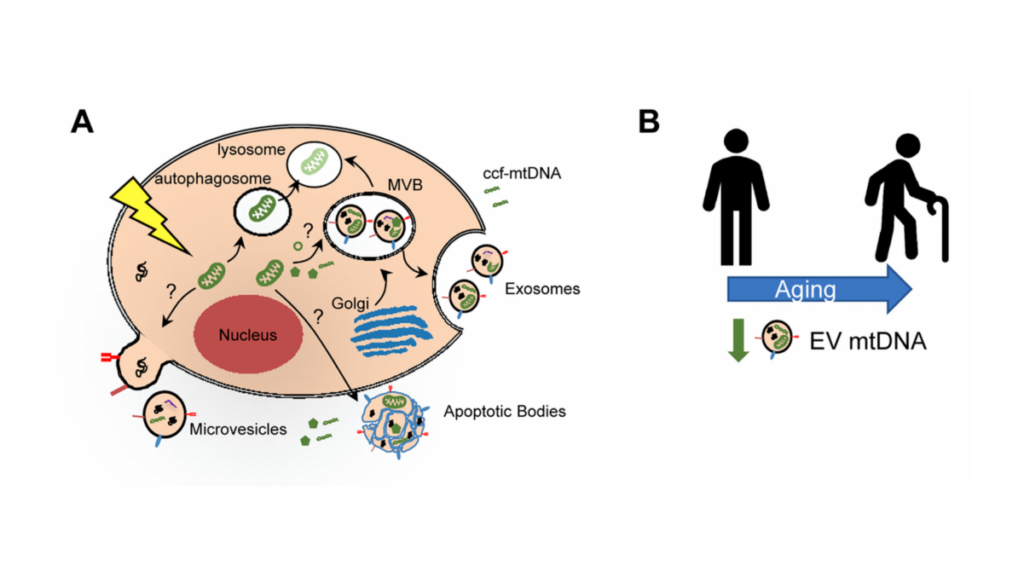
Figure 1. Mitochondrial DNA in extracellular vesicles and association with human aging.
Listen to an audio version of this article

The Trending With Impact series highlights Aging publications that attract higher visibility among readers around the world online, in the news, and on social media—beyond normal readership levels. Look for future science news about the latest trending publications here, and at Aging-US.com.

Some structures inside cells play elusive, yet important roles in human aging. Researchers believe structures classified as extracellular vesicles (EVs), released outside of cell walls, may also play key components in the aging process—specifically related to chronic inflammation.

“EVs are lipid-bound nano-sized vesicles that are secreted outside of cells into the circulation (Figure 1A).”

Two researchers from the National Institutes of Health‘s National Institute on Aging wrote a trending editorial paper, published by Aging in 2021, and entitled, “Mitochondria as extracellular vesicle cargo in aging.” 

Inflammation and Aging

The term “inflamm-aging” has been coined to describe the common state of chronic low-grade inflammation associated with aging. Researchers believe that inflammation contributes to many age-related diseases, including cardiovascular disease, diabetes, cancer, and even dementia.

“In fact, inflammation-related diseases account for more than 50% of worldwide deaths, stressing the importance of inflammation in driving age-related disease and mortality [1,2].”

In the elderly, cellular damage and stress (among other causes) may contribute to chronic inflammation, which can initiate a release of mitochondrial damage-associated molecular patterns. This process can initiate cells to release mitochondrial DNA (mtDNA) into the space outside of the cell as circulating cell-free mitochondria DNA (ccf-mtDNA).

“Due to the similarities between mtDNA and bacterial DNA, this release can in turn elicit a sterile inflammatory response through activation of the innate immune system.”

Circulating Cell-Free Mitochondria DNA

Authors of this editorial believe that ccf-mtDNA may contribute to systemic chronic inflammation. In a previous study, researchers found, in general, that higher plasma/serum levels of ccf-mtDNA were reported in patients with inflammatory-related diseases and after acute injury or infection. However, ccf-mtDNA’s role in aging is complex, as one study showed that ccf-mtDNA levels initially decline into middle-age, and then gradually increase after age 50. 

The molecular details of how ccf-mtDNA exists within blood circulation has yet to be elucidated. Questions still linger surrounding whether or not components in the blood bind to ccf-mtDNA. If components do bind to ccf-mtDNA, are they capable of protecting ccf-mtDNA from destruction in circulation?

Extracellular Vesicles and mtDNA

“Given these gaps in the field, we recently explored whether plasma mtDNA can be encapsulated in extracellular vesicles (EVs) [5].”

The researchers evaluated multiple studies to find that mtDNA can be encapsulated in EVs isolated from plasma—both in cells that have been grown in vitro and in plasma EVs from patients with breast cancer. The next question the researchers addressed was: How do levels of mtDNA in plasma EVs fair in normal conditions and with age?

“To address this need, we isolated plasma EVs and analyzed mtDNA levels with human age. Individuals in this aging cohort had donated plasma at two different time points approximately 5 years apart, which enabled us to examine both crosssectional and longitudinal changes.”

Conclusion

“In both our cross-sectional and longitudinal analyses, EV mtDNA levels decreased with advancing age [5] (Figure 1B).”

The researchers concluded by reporting EV mtDNA levels decreased over a span of five years in the longitudinal cohort. Mitochondrial components, including mtDNA, may be important EV cargo. They emphasized that further research is needed and that it is important for researchers to consider age when using EVs as diagnostic or prognostic markers of disease. 

Click here to read the full editorial paper, published by Aging.

WATCH: MORE AGING VIDEOS ON LABTUBE

Aging is an open-access journal that publishes research papers monthly in all fields of aging research and other topics. These papers are available to read at no cost to readers on Aging-us.com. Open-access journals offer information that has the potential to benefit our societies from the inside out and may be shared with friends, neighbors, colleagues, and other researchers, far and wide.

2021 Ride for Roswell

For media inquiries, please contact media@impactjournals.com.

  • Follow Us