New Insights Into the Mechanisms of Sarcopenia

In this new study, researchers aimed to further elucidate the mechanisms of sarcopenia by examining the influence of denervation in young and middle-aged mice.

New Insights Into the Mechanisms of Sarcopenia

Listen to an audio version of this article

The Trending With Impact series highlights Aging publications (listed as “Aging (Albany NY)” by Medline/PubMed and “Aging-US” by Web of Science) that attract higher visibility among readers around the world online, in the news, and on social media—beyond normal readership levels. Look for future science news about the latest trending publications here, and at Aging-US.com.

A hallmark characteristic of aging is the progressive loss of skeletal muscle mass, known as sarcopenia. A process called motor neuron denervation (Den)—when nerve signals to muscles are blocked or reduced—leads to muscle atrophy, fatigue and eventually muscle loss. Determining how and when Den events influence older muscles is crucially important for developing interventions to stop or reverse age-related muscle wasting.

“Further, aged muscle exhibits reduced plasticity to both enhanced and suppressed contractile activity. It remains unclear when the onset of this blunted response occurs, and how middle-aged muscle adapts to denervation.”

Dysfunctional mitochondria in muscle tissue are known to increase with age. Lysosomes are responsible for the recycling of damaged mitochondria. However, as muscles age, lysosomal function in muscle tissue also declines.

In a new study, researchers Matthew Triolo, Debasmita Bhattacharya and David A. Hood from York University in Toronto, Canada, aimed to characterize the time-dependent changes in denervated skeletal muscle from middle-aged mice. The team focussed on how mitochondrial turnover is impacted. On November 4, 2022, their research paper was published in Aging’s Volume 14, Issue 22, entitled, “Denervation induces mitochondrial decline and exacerbates lysosome dysfunction in middle-aged mice.”

The Study

“The purpose of this study was to compare mitochondrial turnover pathways in young (Y, ~5months) and middle-aged (MA, ~15months) mice, and determine the influence of Den.”

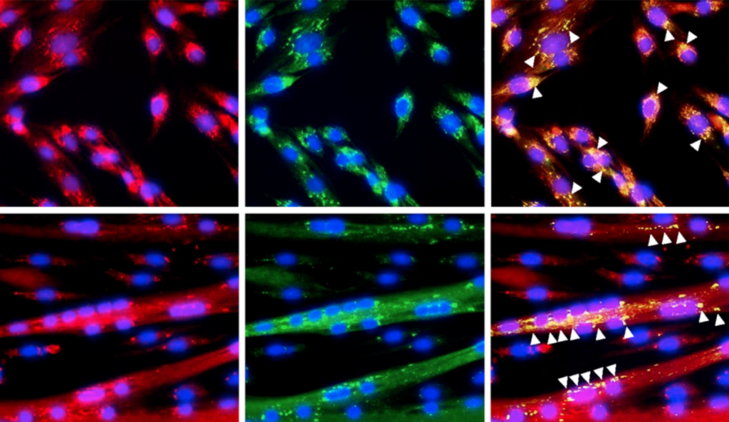
Male mt-Keima mice aged 4-6 months (young) and 14-16 months (middle-aged) were included in this study. The researchers performed surgical procedures to induce Den in the hindlimb muscles of the study mice. After one, three, or seven days of Den, tissue was excised and imaged using confocal microscopy. The researchers collected whole-muscle protein extracts and conducted Western blotting. Statistical analysis was performed using the data they collected.

The middle-aged muscles were compared to muscles from control and young mice. The researchers found that muscle mass, mitochondrial content and PGC-1α protein levels were not different between the young and middle-aged mice. However, indications of enhanced mitochondrial fission and mitophagy and a greater abundance of lysosome proteins were evident in the middle-aged muscle. Their data suggest that increases in fission drive an acceleration of mitophagy in middle-aged murine muscle in order to preserve mitochondrial quality. 

“Den exacerbates the aging phenotype by reducing biogenesis in the absence of a change in mitophagy, perhaps limited by lysosomal capacity, leading to an accumulation of dysfunctional mitochondria with an age-related loss of neuromuscular innervation.”

Conclusion

“In our present study, the inability to upregulate mitophagy flux with denervation is driven by a combination of 1) failure to increase mitophagic proteins and 2) the appearance of dysfunctional lysosomes.”

This latest study may shed light on how muscles age and reveal the importance of mitophagy and lysosomal function in maintaining healthy muscles among middle-aged mice. The study also highlights that denervation induces mitochondrial decline and exacerbates lysosome dysfunction in muscles, thereby worsening age-related muscular atrophy. Further studies are needed to gain a deeper understanding of the mechanisms behind these changes and how they can be prevented or reversed.

“Thus, therapies to combat muscle wasting with age-related physiologic denervation must be designed accordingly. Our results imply targeting both mitochondrial biogenesis and maintenance of lysosome capacity will serve to restore mitochondrial homeostasis and likely metabolic capacity of skeletal muscle.”

Click here to read the full research paper published by Aging.

AGING (AGING-US) VIDEOS: YouTube | LabTube | Aging-US.com

Aging is an open-access journal that publishes research papers bi-monthly in all fields of aging research. These papers are available at no cost to readers on Aging-us.com. Open-access journals have the power to benefit humanity from the inside out by rapidly disseminating information that may be freely shared with researchers, colleagues, family, and friends around the world.

For media inquiries, please contact media@impactjournals.com.

Protein Linked to Aging-Related Muscle Loss

Researchers investigated the mitochondrial protein GRSF1 for its role in the physiology of skeletal muscle aging.

Figure 1. Expression of GRSF1 across myogenesis. (A) RT-qPCR analysis of GRSF1 mRNA levels in proliferating (0 h) and differentiating (24-120 h) human myoblasts; n=3. GRSF1 mRNA levels were normalized to the levels of GAPDH mRNA. (B) Western blot analysis of the levels of GRSF1 at the indicated times during differentiation; n=2. (C) Immunofluorescence detection of GRSF1 (green) and mitochondria (red) in proliferating myoblasts and differentiating myotubes. Arrowheads indicate GRSF1 signals; n=3. Scale bar, 50 μm.
Listen to an audio version of this article

Skeletal muscle is responsible for regulating physical movement and comprises between 30 and 40% of the human body’s mass. The loss of skeletal muscle has major impacts on overall health and quality of life—leading to frailty and a decreased ability to perform activities of daily living. The most common cause of muscle loss is aging, and a prevalent pattern of aging-associated muscular decline is known as sarcopenia.

“With advancing age, the progressive loss of skeletal muscle mass and function, known as sarcopenia, leads to reduced muscle strength and diminishes individual mobility, quality of life, and lifespan [12].”

In a research paper published in Aging (Aging-US) Volume 13, Issue 11, researchers from the National Institutes of Health’s National Institute on Aging and Chungnam National University investigated a protein that may play a role in aging-related muscle loss. Their paper was published on June 2, 2021, and entitled, “GRSF1 deficiency in skeletal muscle reduces endurance in aged mice.”

Skeletal Muscles and Mitochondrial Proteins

The healthy operation of skeletal muscle is dependent on well-regulated mitochondrial functioning. Skeletal muscle is extremely rich in mitochondria, as mitochondria supply muscle cells with the energy they need to help move the body, known as adenosine 5′-triphosphate, or ATP. With age, the mitochondria in skeletal muscles begin to progressively malfunction. The exact mechanisms involved in this decline have not been fully elucidated.

“In aging skeletal muscle, mitochondria display reduced function, altered morphology, and increased production of reactive oxygen species (ROS), which contribute to a progressive loss of muscle mass and strength [1314].”

The guanine-rich RNA sequence binding factor 1 (GRSF1) protein is widely distributed in mammalian organs, and primarily enriched in mitochondria organelles. This makes the skeletal muscle an ideal organ in which researchers can study GRSF1, and other mitochondrial proteins, to investigate their role in aging-related processes such as sarcopenia. Although GRSF1 has been well-studied for its role in maintaining mitochondrial function, the involvement of GRSF1 in skeletal muscle aging had not yet been investigated until this study.

The Study

In this study, the researchers used Grsf1cKO mice—a mouse model in which GRSF1 is specifically knocked out in murine skeletal muscle cells. The mice appeared normal until 7-9 months of age. At 16-18 months of age, however, the researchers observed a reduction in muscle endurance compared to wild-type (WT) control mice. The authors postulated that these results suggested the loss of GRSF1 in skeletal muscle may not alter muscle function until later in life.

“The Grsf1cKO mice at this age ran about a 30% shorter treadmill distance on average relative to WT controls (Figure 3A).”

Upon further transcriptomic analysis, the team found that more than 200 muscle genes were differentially expressed in the GRSF1-deficient mice compared to the control mice. Some of the differentially expressed RNAs that were elevated in the Grsf1cKO mice were the hypoxia-inducible Mgarp mRNA, the mRNA encoding Sarcolipin (SLN), the pro-inflammatory proteins CXCL10 and NFKB2, and the transcription factor ATF3. The authors suggested that increased SLN mRNA may also potentially contribute to the decline in skeletal muscle endurance seen in Grsf1cKO mice.

“The reduction of endurance in Grsf1cKO muscle was accompanied by differential expression of several mRNAs, including some that encoded mitochondrial proteins, inflammatory proteins, ion transporters, and transcription factors (Mgarp, Sln, Cxcl10, Nfkb2, and Atf3 mRNAs).”

Conclusion

The researchers found that the absence of GRSF1 in murine skeletal muscle cells led to a decrease in muscle endurance. Initially, the researchers had anticipated that GRSF1 knock-out would lead to a dramatic loss in muscle function. However, their study revealed that the function of GRSF1 in skeletal muscle appeared to only be moderate. Overall, this is an important finding, as it provides new insights into the role of GRSF1 in muscle physiology and opens up new avenues for research into potential therapies for aging-related muscle loss.

“This modest in vivo effect suggests that there are redundant or compensatory mechanisms that prevent catastrophic damage from GRSF1 loss in aging muscle, and that identifying such factors might be of therapeutic benefit in diseases caused by impaired function of muscle mitochondria and impaired muscle regeneration.”

Click here to read the full priority research paper published in Aging (Aging-US).

AGING (AGING-US) VIDEOS: YouTube | LabTube | Aging-US.com

Aging (Aging-US) is an open-access journal that publishes research papers bi-monthly in all fields of aging research. These papers are available at no cost to readers on Aging-us.com. Open-access journals have the power to benefit humanity from the inside out by rapidly disseminating information that may be freely shared with researchers, colleagues, family, and friends around the world.

For media inquiries, please contact media@impactjournals.com.

Trending With Impact: Are Our Muscles Intrinsically Impaired by Aging?

In a priority research paper published by Aging-US in January of 2022, researchers investigated aged muscle stem cells and their ability to sense and respond to mechanical cues.

Listen to an audio version of this article
3D Illustration of muscle tissue

The Trending With Impact series highlights Aging (Aging-US) publications that attract higher visibility among readers around the world online, in the news, and on social media—beyond normal readership levels. Look for future science news about the latest trending publications here, and at Aging-US.com.

IIs muscle wasting a fate humans can avoid, or will the problem of aging-related muscle loss only be resolved when the mystery of aging is solved? Researchers—from Vrije Universiteit AmsterdamUniversity of AmsterdamSorbonne UniversitéAmsterdam University Medical Center VUmcUniversité Catholique de LouvainKU Leuven, and Institut NeuroMyoGène—conducted a study aimed at elucidating whether muscle stem cells are inherently impaired by the aging process in their ability to sense and respond to mechanical cues. Their priority research paper was published in January of 2022 on the cover of Aging (Aging-US) Volume 14, Issue 1, and entitled, “Reduced growth rate of aged muscle stem cells is associated with impaired mechanosensitivity.”

Muscle Stem Cells

Muscle stem cells (MuSCs) are stem cells located within skeletal muscle tissues. MuSCs function to repair Muscle stem cells (MuSCs) are stem cells located within skeletal muscle tissues. MuSCs function to repair damaged myofibers and give rise to new skeletal muscle cells. These self-renewing stem cells are involved in muscle growth, repair and regeneration. As we age, MuSCs decline in number and lose their potential to regenerate damaged myofibers, leading to sarcopenia. The researchers in this study hypothesized that the responsiveness of aged MuSCs is impared by the aging process both physically and mechanically.

“We postulated that aged MuSCs are intrinsically impaired in their responsiveness to omnipresent mechanical cues through alterations in MuSC morphology, mechanical properties, and number of integrins, culminating in impaired proliferative capacity.”

The Study

The researchers assessed whether aged MuSCs become impaired in their ability to proliferate, respond to pulsating fluid shear stress (PFSS) mechanical loading, maintain focal adhesion number and/or size after mechanical loading, and in their ability to express the protein-coding gene Integrin Subunit Alpha 7 (ITGA7). 

“Integrins are transmembrane protein receptors that connect MuSCs to the ECM [extracellular matrix] components and are part of focal adhesions [51].”

Young MuSCs (2 months) and aged MuSCs (22 months) were isolated from male mice. Fluorescence-activated cell purification was carried out and cells were cultured. To measure proliferation, images were captured of the cell cultures every 24 hours. Images were also taken pre- and post-PFSS to determine the number of young and aged MuSCs detached from the culture media (focal adhesion) as a result of PFSS treatment. Since nitric oxide (NO) is known to play a role in MuSC activation and muscle regeneration, NO analysis was conducted to measure NO production. To determine MuSC morphology, the researchers carried out immunohistochemistry staining. They also measured MuSC stiffness, deformation, gene expression, and RNA isolation and reverse transcription.

Compared to young MuSCs, the researchers found aged MuSCs had impaired growth. Their results showed that IL-6 gene expression was lower in aged MuSCs, which suggested that aged MuSCs were intrinsically altered in the signaling pathways governing proliferation and MuSC function. Aged MuSCs showed an increase in cell volume and reduced cell adhesion after mechanical loading. NO levels in young and aged MuSCs were similar, and PFSS in both cultures resulted in similar increases in NO production. The researchers found decreased ITGA7 expression and reduced pPXN clusters (focal adhesion formation) were involved in altered MuSC function with age. High YAP nuclear localization was found in aged MuSCs, as well as reduced mechanosensitivity.

“Aged MuSCs were less sensitive to shear forces and showed upregulation of less genes, suggesting that the decreased mechanosensitivity was due to decreased integrin protein expression, i.e. ITGA7, ITGA5, and ITGB5, and focal adhesion number.”

Conclusion

The results from this study found that aged MuSCs were intrinsically impaired in their growth rate due to decreased ITGA7 expression and diminished focal adhesion formation. These changes coincided with increased cell volume, decreased MuSC adhesion, altered mechanosensitivity, changed YAP signaling and decreased expression of several genes (including cell cycle genes). The researchers suggest that ITGA7 and pPXN may be potential therapeutic targets to improve aged MuSC function.

“As an implication, a possible therapeutic option could be restoration ITGA7 and focal adhesion number in aged MuSCs, which may help to restore MuSCs adhesion to their niche as well as growth rate of these cells.”

Click here to read the full priority research paper published by Aging (Aging-US).

AGING (AGING-US) VIDEOS: YouTube | LabTube | Aging-US.com

Aging (Aging-US) is an open-access journal that publishes research papers bi-monthly in all fields of aging research. These papers are available to read at no cost to readers on Aging-us.com. Open-access journals offer information that has the potential to benefit our societies from the inside out and may be shared with friends, neighbors, colleagues, and other researchers, far and wide.

For media inquiries, please contact media@impactjournals.com.

  • Follow Us