In this new study, researchers investigated the senescent phenotypes of human corneal endothelial cells upon UV-A exposure.
—
With an ever-increasing global population grappling with age-related ocular ailments like cataracts, dry eyes, glaucoma, and macular degeneration, the need for new research in this domain is more pressing than ever.
In a new study, researchers Kohsaku Numa, Sandip Kumar Patel, Zhixin A. Zhang, Jordan B. Burton, Akifumi Matsumoto, Jun-Wei B. Hughes, Chie Sotozono, Birgit Schilling, Pierre-Yves Desprez, Judith Campisi (1948-2024), and Koji Kitazawa from the Buck Institute for Research on Aging, Kyoto Prefectural University of Medicine, University of Cambridge, and California Pacific Medical Center shed light on a pivotal aspect of corneal health – the impact of ultraviolet-A (UV-A) radiation on corneal endothelial cells. Their research paper was published on the cover of Aging’s Volume 16, Issue 8, entitled, “Senescent characteristics of human corneal endothelial cells upon ultraviolet-A exposure.”
“The objective of this study was to investigate the senescent phenotypes of human corneal endothelial cells (hCEnCs) upon treatment with ultraviolet (UV)-A.”
Corneal Health & Cellular Senescence
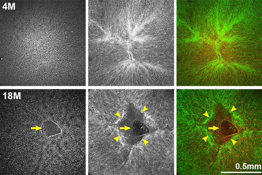
The cornea, a transparent tissue responsible for refracting incoming light onto the retina, plays a crucial role in our visual acuity. Its transparency is maintained by a single layer of cells called corneal endothelial cells (CEnCs), which cover the posterior surface. However, these cells possess a limited capacity for proliferation, rendering them susceptible to pathological cell loss, potentially leading to corneal endothelial dysfunction and, ultimately, visual impairment or blindness.
Current treatments for CEnC dysfunction include corneal endothelial transplantation using donor corneas and cell injection therapy utilizing cultured human CEnCs (hCEnCs). Nonetheless, pathological CEnC loss persists even after successful interventions, culminating in graft failure. To combat this, researchers have delved into the intricate mechanisms underlying hCEnCs loss, uncovering a potential link between corneal endothelial disease and cellular senescence.
While cellular senescence acts as a natural defense mechanism against uncontrolled cell proliferation, the accumulation of senescent cells can exacerbate pathological conditions and contribute to various age-related etiologies. Notably, senescent cells acquire an inflammatory phenotype known as the senescence-associated secretory phenotype (SASP), which can adversely alter the surrounding microenvironment over time.
The Study
In the current study, the researchers exposed hCEnCs to varying doses of UV-A radiation, ranging from 0 J/cm2 (mock) to 20 J/cm2. Cells treated with 10 Gy of ionizing radiation (IR) served as positive controls for senescence induction.
“UV-A accounts for about 90% of the UV radiation reaching the earth’s surface and is known to induce ROS causing oxidative stress [34]. Oxidative stress causes molecular alternation, leading to cellular senescence [35]. Observations of UV-A intensity suggest that exposure to 5 J/cm2 of UV-A is roughly equivalent to one hour of noonday sun exposure during the summer [34].”
Through a meticulous analysis of cell morphology, senescence-associated β-galactosidase (SA-β-gal) activity, cell proliferation, and expression of senescence markers (p16 and p21), the team identified that hCEnCs exposed to 5 J/cm2 of UV-A exhibited typical senescent phenotypes, including enlargement, increased SA-β-gal activity, decreased cell proliferation, and elevated expression of p16 and p21. The researchers employed RNA sequencing (RNA-Seq) and proteomics analysis to gain a comprehensive understanding of the senescence response in hCEnCs.
Results
RNA-Seq analysis revealed a significant overlap in the pathways modulated by UV-A and IR-induced senescence. Upregulated genes were enriched in pathways associated with extracellular matrix (ECM) organization, cellular component movement, response to cytokines, cell migration, and motility – processes intimately linked to corneal endothelial diseases.
Interestingly, while the number of significantly up- or down-regulated genes differed between UV-A and IR exposure, the proteomics analysis revealed a much smaller disparity in the number of altered proteins, suggesting that UV-A might be a more physiologically relevant method for inducing cellular senescence in hCEnCs. The proteomics analysis unveiled a wealth of information regarding the SASP of UV-A-induced senescent hCEnCs. Key SASP components, including STC1, GDF15, C7, C9, SERPINE2, and PDGFA, were identified among the top 40 secreted proteins.
The researchers also detected elevated levels of CXCL1, CXCL8, MMP2, COL6A2, COL8A1, COL12A1, and other proteins previously reported as SASP factors in various cell types. Notably, proteins associated with glycolysis, such as SLC2A1, GPI, ENO1, PKM, TPI1, and LDH, were also found to be significantly upregulated.
Conclusions & Future Directions
“Here, we showed that cellular senescence is induced in hCEnCs upon UV-A irradiation and conducted comprehensive analyses of RNA and protein expression.”
This study not only sheds light on the senescent characteristics of hCEnCs upon UV-A exposure but also highlights the potential role of cellular senescence in the pathogenesis of corneal endothelial diseases. By identifying the overlapping pathways and SASP factors modulated by both UV-A and IR-induced senescence, the researchers have paved the way for a deeper understanding of the molecular mechanisms underlying CEnC dysfunction.
Furthermore, the identification of specific proteins associated with corneal endothelial diseases, such as TGFBI, TGFB1, TGFB2, LOXL1, LOXL2, and complement factors, provides valuable insights into potential therapeutic targets and biomarkers for early detection and intervention.
As the research community continues to unravel the enigma of cellular senescence and its implications in ocular health, this study stands as a testament to the power of multidisciplinary approaches and cutting-edge techniques in advancing our understanding of age-related vision impairment.
Click here to read the full research paper published in Aging.
—
Aging is an open-access, traditional, peer-reviewed journal that publishes high-impact papers in all fields of aging research. All papers are available to readers (at no cost and free of subscription barriers) in bi-monthly issues at Aging-US.com.
Click here to subscribe to Aging publication updates.
For media inquiries, please contact [email protected].