“This collection is published in memory of Professor Judith Campisi, a pioneering force in the field of cellular senescence whose groundbreaking work shaped the understanding of senescence in aging, cancer, and tissue homeostasis.”
BUFFALO, NY — May 1, 2025 —Aging (Aging-US) invites submissions for a Special Collection dedicated to the theme of cellular senescence, spanning its basic mechanisms, physiological and pathological functions, and clinical applications.
This collection is published in memory of Professor Judith Campisi, a pioneering force in the field of cellular senescence whose groundbreaking work shaped the understanding of senescence in aging, cancer, and tissue homeostasis. Her legacy continues to inspire generations of scientists working to decode the complex biology of senescent cells and their impact on health and disease.
We welcome original research articles, reviews, and perspectives on topics including:
Fundamental mechanisms of senescence induction and maintenance
Regulation and context-specific roles of the senescence-associated secretory phenotype (SASP)
Beneficial and detrimental effects of senescent cells in vivo
Senescence in development, aging, regeneration, and age-related diseases
Biomarkers, imaging, and tools for senescence detection and quantification
Therapeutic targeting of senescent cells: senolytics, senomorphics, and clinical translation
This Special Collection is guest edited by Han Li and Irina Conboy, both internationally recognized leaders in the study of senescence and aging.
In a new study, researchers investigated myocyte-secreted factors with the potential to suppress cellular senescence, aiming to explore their protective effects against lung disease.
—
Over the human lifespan, our cells encounter numerous stressors that can trigger an intrinsic defense mechanism called cellular senescence. Cellular senescence is characterized by irreversible growth arrest and can act as a safeguard against cancer. However, when senescent cells accumulate in various tissues as we age, it can contribute to tissue degeneration and chronic diseases.
The senescence-associated secretory phenotype (SASP), a hallmark of senescent cells, plays a critical role by secreting inflammatory factors, proteases, and growth factors, disrupting tissue balance and fueling pathological conditions. Consequently, selectively eliminating senescent cells has emerged as a promising therapeutic strategy, potentially restoring tissue function and mitigating age-related disorders.
COPD: Chronic Obstructive Pulmonary Disease
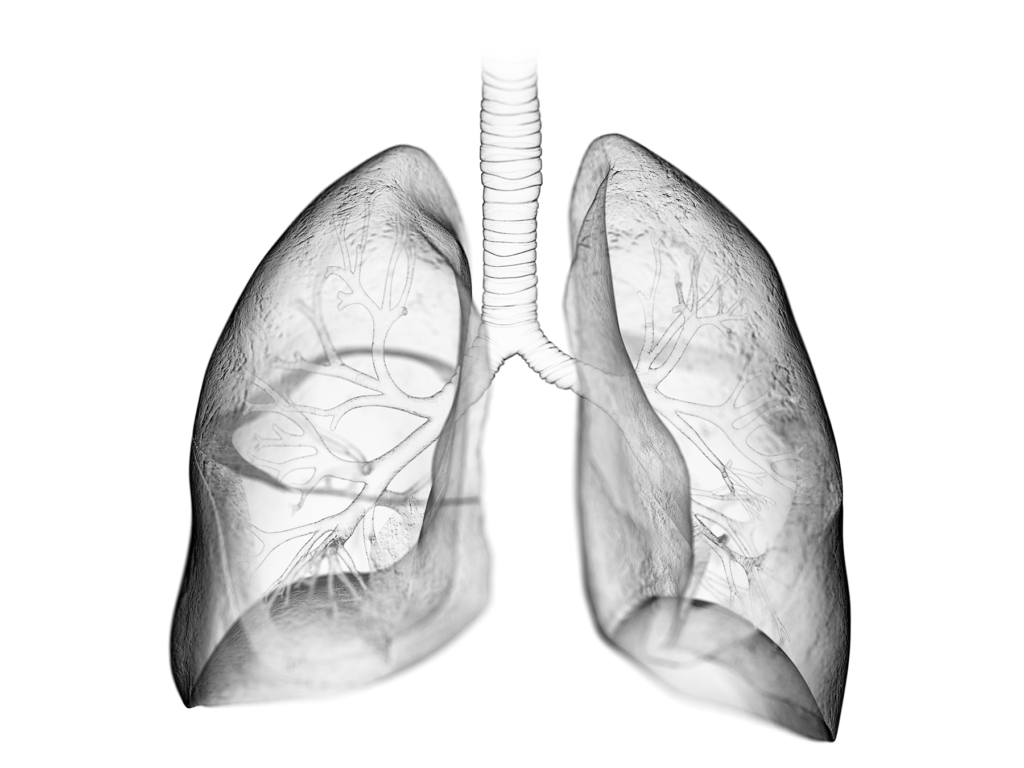
Chronic obstructive pulmonary disease (COPD) exemplifies the impact of cellular senescence on health, characterized by the collapse of alveolar walls in the lungs. Accelerated accumulation of senescent cells in COPD patients’ lung tissues links senescence to the disease’s pathogenesis. Genetic or pharmacological elimination of these cells in preclinical models has shown significant reductions in emphysema-associated pathologies and restoration of pulmonary function, highlighting the potential of senolytic therapies.
Regular physical activity offers benefits beyond fitness, including cardiovascular and mental well-being enhancements, and modulates cellular senescence. Studies show an association between habitual exercise and lower levels of senescence markers in various tissues. Researchers have focused on myokines, signaling factors secreted by skeletal muscles in response to exercise, as potential mediators of these benefits. Irisin, a myokine, has shown promise in suppressing cellular senescence and correlating inversely with COPD severity.
In this recent study, pigment epithelium-derived factor (PEDF) emerged as a key player in the interplay between exercise, cellular senescence, and lung pathologies. Initially known for its role in retinal development, PEDF has been linked to cellular senescence modulation, extending the replicative lifespan of fibroblasts and diminishing senescence markers. PEDF mitigates oxidative stress by reducing reactive oxygen species levels and modulates microRNAs, particularly miR-127, implicated in cellular senescence.
“We found that myocyte-derived factors significantly extended the replicative lifespan of fibroblasts, suggesting that myokines mediate the anti-senescence effects of exercise.”
Exercise significantly upregulates PEDF expression in skeletal muscles, correlating with reduced senescence markers and SASP-related genes in the lungs. Recombinant PEDF administration in mice has shown remarkable results, reducing senescence markers and preserving alveolar structure in pulmonary emphysema models, translating into improved pulmonary function. While some preclinical evidence supports PEDF’s therapeutic potential, translating these findings to clinical applications requires rigorous safety and efficacy evaluations. Understanding PEDF’s signaling pathways could unveil new therapeutic targets, and its potential involvement in other age-related disorders warrants further investigation. The interplay between PEDF and other exercise-induced factors offers potential for novel therapeutic strategies.
“Collectively, these results strongly suggest that PEDF contributes to the beneficial effects of exercise, potentially suppressing cellular senescence and its associated pathologies.”
Conclusions
The discovery of PEDF’s role in exercise-induced senescence suppression and its therapeutic potential in lung pathologies represents a paradigm shift in senescence research. Understanding the interplay between physical activity, myokine signaling, and senescence modulation can lead to targeted interventions promoting healthy aging. Multidisciplinary collaborations are essential to harness the potential of PEDF and other senescence-modulating factors, paving the way for innovative treatments that alleviate age-related diseases and improve quality of life.
Aging is an open-access, traditional, peer-reviewed journal that publishes high-impact papers in all fields of aging research. All papers are available to readers (at no cost and free of subscription barriers) in bi-monthly issues at Aging-US.com.
Click here to subscribe to Aging publication updates.
In this new study, researchers investigated the senescent phenotypes of human corneal endothelial cells upon UV-A exposure.
—
With an ever-increasing global population grappling with age-related ocular ailments like cataracts, dry eyes, glaucoma, and macular degeneration, the need for new research in this domain is more pressing than ever.
“The objective of this study was to investigate the senescent phenotypes of human corneal endothelial cells (hCEnCs) upon treatment with ultraviolet (UV)-A.”
Corneal Health & Cellular Senescence
The cornea, a transparent tissue responsible for refracting incoming light onto the retina, plays a crucial role in our visual acuity. Its transparency is maintained by a single layer of cells called corneal endothelial cells (CEnCs), which cover the posterior surface. However, these cells possess a limited capacity for proliferation, rendering them susceptible to pathological cell loss, potentially leading to corneal endothelial dysfunction and, ultimately, visual impairment or blindness.
Current treatments for CEnC dysfunction include corneal endothelial transplantation using donor corneas and cell injection therapy utilizing cultured human CEnCs (hCEnCs). Nonetheless, pathological CEnC loss persists even after successful interventions, culminating in graft failure. To combat this, researchers have delved into the intricate mechanisms underlying hCEnCs loss, uncovering a potential link between corneal endothelial disease and cellular senescence.
While cellular senescence acts as a natural defense mechanism against uncontrolled cell proliferation, the accumulation of senescent cells can exacerbate pathological conditions and contribute to various age-related etiologies. Notably, senescent cells acquire an inflammatory phenotype known as the senescence-associated secretory phenotype (SASP), which can adversely alter the surrounding microenvironment over time.
The Study
In the current study, the researchers exposed hCEnCs to varying doses of UV-A radiation, ranging from 0 J/cm2 (mock) to 20 J/cm2. Cells treated with 10 Gy of ionizing radiation (IR) served as positive controls for senescence induction.
“UV-A accounts for about 90% of the UV radiation reaching the earth’s surface and is known to induce ROS causing oxidative stress [34]. Oxidative stress causes molecular alternation, leading to cellular senescence [35]. Observations of UV-A intensity suggest that exposure to 5 J/cm2 of UV-A is roughly equivalent to one hour of noonday sun exposure during the summer [34].”
Through a meticulous analysis of cell morphology, senescence-associated β-galactosidase (SA-β-gal) activity, cell proliferation, and expression of senescence markers (p16 and p21), the team identified that hCEnCs exposed to 5 J/cm2 of UV-A exhibited typical senescent phenotypes, including enlargement, increased SA-β-gal activity, decreased cell proliferation, and elevated expression of p16 and p21. The researchers employed RNA sequencing (RNA-Seq) and proteomics analysis to gain a comprehensive understanding of the senescence response in hCEnCs.
Results
RNA-Seq analysis revealed a significant overlap in the pathways modulated by UV-A and IR-induced senescence. Upregulated genes were enriched in pathways associated with extracellular matrix (ECM) organization, cellular component movement, response to cytokines, cell migration, and motility – processes intimately linked to corneal endothelial diseases.
Interestingly, while the number of significantly up- or down-regulated genes differed between UV-A and IR exposure, the proteomics analysis revealed a much smaller disparity in the number of altered proteins, suggesting that UV-A might be a more physiologically relevant method for inducing cellular senescence in hCEnCs. The proteomics analysis unveiled a wealth of information regarding the SASP of UV-A-induced senescent hCEnCs. Key SASP components, including STC1, GDF15, C7, C9, SERPINE2, and PDGFA, were identified among the top 40 secreted proteins.
The researchers also detected elevated levels of CXCL1, CXCL8, MMP2, COL6A2, COL8A1, COL12A1, and other proteins previously reported as SASP factors in various cell types. Notably, proteins associated with glycolysis, such as SLC2A1, GPI, ENO1, PKM, TPI1, and LDH, were also found to be significantly upregulated.
Conclusions & Future Directions
“Here, we showed that cellular senescence is induced in hCEnCs upon UV-A irradiation and conducted comprehensive analyses of RNA and protein expression.”
This study not only sheds light on the senescent characteristics of hCEnCs upon UV-A exposure but also highlights the potential role of cellular senescence in the pathogenesis of corneal endothelial diseases. By identifying the overlapping pathways and SASP factors modulated by both UV-A and IR-induced senescence, the researchers have paved the way for a deeper understanding of the molecular mechanisms underlying CEnC dysfunction.
Furthermore, the identification of specific proteins associated with corneal endothelial diseases, such as TGFBI, TGFB1, TGFB2, LOXL1, LOXL2, and complement factors, provides valuable insights into potential therapeutic targets and biomarkers for early detection and intervention.
As the research community continues to unravel the enigma of cellular senescence and its implications in ocular health, this study stands as a testament to the power of multidisciplinary approaches and cutting-edge techniques in advancing our understanding of age-related vision impairment.
Click here to read the full research paper published in Aging.
—
Aging is an open-access, traditional, peer-reviewed journal that publishes high-impact papers in all fields of aging research. All papers are available to readers (at no cost and free of subscription barriers) in bi-monthly issues at Aging-US.com.
Click here to subscribe to Aging publication updates.
In a new study, researchers aimed to investigate the prognostic significance of senescence-related TME genes in head and neck squamous cell carcinoma (HNSCC) and their potential implications for immunotherapy response.
—
Head and neck squamous cell carcinoma (HNSCC) is a prevalent and heterogeneous form of cancer that affects thousands of individuals worldwide. The prognosis for HNSCC patients can vary greatly, depending on factors such as tumor stage and site. The tumor microenvironment (TME) plays a crucial role in tumorigenesis and disease progression, with cellular senescence being a key component. Senescent cells, characterized by cell-cycle arrest, have been shown to have both tumor-suppressive and tumor-promoting effects. However, the prognostic significance of senescence-related TME genes in HNSCC remains poorly understood.
In a new study, researchers Young Chan Lee, Yonghyun Nam, Minjeong Kim, Su Il Kim, Jung-Woo Lee, Young-Gyu Eun, and Dokyoon Kim from Kyung Hee University, Kyung Hee University Hospital at Gangdong, and the University of Pennsylvania aimed to investigate the prognostic significance of senescence-related TME genes in HNSCC and their potential implications for immunotherapy response. They utilized data from The Cancer Genome Atlas (TCGA) to identify two distinct subtypes of HNSCC based on the expression of senescence-related TME genes. The team then constructed a risk model consisting of senescence-related TME core genes (STCGs) and validated its prognostic capability in independent cohorts. Their research paper was chosen as an Aging cover paper and published in Volume 16, Issue 2, entitled, “Prognostic significance of senescence-related tumor microenvironment genes in head and neck squamous cell carcinoma.”
“To the best of our knowledge, this is the first study to offer a comprehensive evaluation of the senescence related TME status by integrating senescence related TME genes through a gene-gene network and clustering. Furthermore, we have introduced a novel risk model that utilizes a selected gene set to predict prognosis and confirmed the expression of STCGs in immune cells at single-cell levels.”
The Study
Identification of Prognostic Senescence-Related TME Genes
To identify prognostic senescence-related TME genes, the researchers screened a total of 7,878 genes in the TCGA-HNSCC dataset. They identified 288 genes that belonged to TME-related genes, tumor-associated senescence (TAS) genes, and immune-related genes. From these genes, they selected 91 prognostic senescence-related TME genes (PSTGs) based on differential expression analysis and Cox regression analysis.
Senescence-Related TME Subtypes and Characterization
Using consensus clustering analysis, the researchers classified the HNSCC samples into two distinct subtypes based on the expression of PSTGs: subtype 1 and subtype 2. The two subtypes exhibited significant differences in clinical and molecular characteristics. Subtype 2 had a higher prevalence of HPV-positive and oropharyngeal cancer cases, while subtype 1 was characterized by a higher proportion of advanced tumor stage and overall stage.
Further analysis revealed distinct differences between the subtypes in terms of genetic alterations, methylation patterns, enriched pathways, and immune infiltration. Subtype 1 had a higher mutation rate in the TP53 gene and exhibited hypomethylation in several CpG sites compared to subtype 2. Additionally, subtype 2 showed higher immune scores, stromal scores, and ESTIMATE scores, indicating a more favorable immune microenvironment.
The two subtypes also displayed differences in survival outcomes. Kaplan-Meier survival analysis showed that subtype 2 had a more favorable overall survival compared to subtype 1. This difference was enhanced in the HPV-positive cohort, suggesting that the senescence-related TME subtypes may have implications for prognosis in specific patient subgroups.
Risk Scoring Based on Senescence-Related TME Status
Using the 91 PSTGs, the researchers constructed a risk scoring model based on the LASSO Cox regression algorithm. They identified 21 STCGs that were associated with either increased risk or protection. The risk scores based on the expression levels of these genes were calculated for each patient, and the patients were classified into high- and low-risk groups.
The prognostic performance of the risk scoring model was tested in independent cohorts, including the TCGA-HNSCC test set, the GSE41613 cohort, and the KHUMC cohort. The high-risk group showed significantly lower overall survival compared to the low-risk group in the TCGA-HNSCC test set and the GSE41613 cohort. Although not statistically significant, the low-risk group demonstrated a trend towards higher overall survival in the KHUMC cohort.
Immunotherapy Response Prediction and Single-Cell Analysis
The team also investigated the immunotherapy response prediction based on the risk model and the expression of STCGs. They found that the low-risk group had higher immunophenoscores and a significantly higher proportion of responders to immunotherapy compared to the high-risk group.
To further evaluate the senescence-related TME characteristics at the single-cell level, the researchers analyzed single-cell transcriptome data from HNSCC tissue. They found that STCGs were enriched in fibroblast, mono/macrophage, and T cell populations, suggesting that these cell types contribute to the senescent features of HNSCC.
Conclusion
In conclusion, the study sheds light on the prognostic significance of senescence-related TME genes in HNSCC. Their findings highlight the heterogeneity of HNSCC and the importance of the senescence-related TME in prognosis and immunotherapy response. The risk scoring model based on STCGs provides a potential prognostic biomarker for HNSCC patients, and the single-cell analysis further elucidates the association between STCGs and specific cell populations within the TME. These findings contribute to a deeper understanding of the complex interplay between senescence and the TME in HNSCC and have implications for precision medicine and personalized treatment approaches. Further research and validation are needed to fully understand the clinical implications of senescence-related TME genes in HNSCC. However, this study provides valuable insights into the role of cellular senescence in tumor progression and the potential for targeting senescence-related pathways in the development of novel therapeutic strategies for HNSCC patients.
“In conclusion, this study comprehensively investigated the prognostic and immunological features of senescence related TME genes in HNSC. By leveraging these senescence related TME genes, we successfully developed a risk model to predict HNSC prognosis and immunotherapy response, which was robustly validated using external transcriptome datasets. These findings provided evidence for the role of senescence in the TME and highlighted the potential of senescence-related biomarkers as promising therapeutic targets.”
Click here to read the full research paper published in Aging.
—
Aging is an open-access, traditional, peer-reviewed journal that has published high-impact papers in all fields of aging research since 2009. All papers are available to readers (at no cost and free of subscription barriers) in bi-monthly issues at Aging-US.com.
Click here to subscribe to Aging publication updates.
In a new study, researchers aimed to reveal a link between telomere dysfunction, ovarian aging and infertility using a mouse model of accelerated-reproductive senescence.
Telomeres are the protective caps at the ends of chromosomes that prevent DNA damage and maintain genomic stability. However, telomeres shorten with each cell division and eventually reach a critical length that triggers cellular senescence or death. Telomere length (TL) and telomerase activity (TA), the enzyme that replenishes telomeric repeats, are influenced by genetic and environmental factors and vary among tissues and individuals.
“Telomere attrition has been identified as one of the molecular determinants of aging [7].”
Telomere dysfunction has been implicated in various age-related diseases, including infertility. Ovarian aging is the main cause of infertility in women, as it leads to a decline in both the quantity and quality of oocytes. Previous studies have shown that TL and TA are reduced in oocytes and granulosa cells of women with diminished ovarian reserve or poor response to ovarian stimulation. Moreover, TL and TA have been associated with ovarian reserve markers and pregnancy outcomes in assisted reproductive technologies.
The SAMP8 mouse model, which has previously been suggested as an Alzheimer’s disease model of aging, also exhibits a shortened estrous cycle, elevated follicle-stimulating hormone (FSH) levels, and reduced fertility in females at just seven months of age. SAMP8 mice have a shorter lifespan compared to senescence-accelerated mouse resistant 1 (SAMR1) mice. SAMR1 mice do not exhibit reproductive senescence. Thus, the researchers deemed the SAMR1 mouse model an appropriate control group to study the SAMP8 mouse model as a model of ovarian aging and infertility.
“In the current study, we sought to investigate whether the SAMP8 mice, which show accelerated-reproductive senescence, have alterations in their telomere pathway. This question has not yet been explored in relation to reproduction in this model.”
In this study, the team compared the TL and TA in blood and ovary samples from the SAMP8 female mice at seven months of age (when they show signs of reproductive senescence) with age-matched control SAMR1 mice. They also evaluated the ovarian follicle development, the expression of telomerase subunits (TERT and TERC), and the reproductive outcomes after ovarian stimulation in both groups of mice. In sum, the researchers measured survival rates (in male and female mice), alteration in the telomere pathway at seven months of age, TERT and TERC expression levels, TA on the TL of granulosa cells in developing follicles, and impairment/alterations in the telomere pathway in oogenesis and embryo development.
The results revealed that SAMP8 females had a reduced median lifespan compared to SAMP8 males and SAMR1 males and females. In blood, SAMP8 females had lower mean TL and higher accumulation of short telomeres than the other mice. In ovary, SAMP8 females had lower TA and TERT expression. Furthermore, SAMP8 females had fewer primordial, primary, secondary, and antral follicles than control females, indicating a diminished ovarian reserve. After ovarian stimulation, SAMP8 females had a lower number of oocytes than controls of the same age. Their results suggested that oogenesis and embryo development is impaired in SAMP8 mice at seven months compared to age-matched controls, and this coincides with alterations in the telomere pathway.
Conclusions
“Thus, SAMP8 females represent a bona fide model for the analysis of fertility, not only because it shows similar phenotype to middle-aged women as stated earlier [43], but also because the alterations in the telomere pathway are found in women with fertility disorders [37, 38, 40, 41] and this pathway links reproduction with longevity.”
The researchers concluded that SAMP8 females have impaired telomere pathway and fertility, reflecting signs of reproductive senescence described in middle-aged women. They suggested that the SAMP8 model could be useful in studying the role of telomere dysfunction in ovarian aging and infertility. In addition, this mouse model could be used to test potential therapeutic interventions to improve female reproductive health.
“Understanding the molecular pathways underlying aging and fertility, provides a basis for further studies focused on several topics. First, the analysis of embryo alterations, which can be better assessed in mice than in humans. Second, how reproductive lifespan improvement may ameliorate elderly health. And third, the mechanisms underlying follicle recruitment and development, which are not completely known.”
Click here to read the full research paper published by Aging.
—
Aging is an open-access, peer-reviewed journal that has been publishing high-impact papers in all fields of aging research since 2009. These papers are available to readers (at no cost and free of subscription barriers) in bi-monthly issues at Aging-US.com.
Click here to subscribe to Aging publication updates.
A new study by researchers from Osaka University’s Graduate School of Dentistry investigated cellular senescence in periodontal tissue and disease—identifying promising therapeutic targets for preventing periodontitis in the elderly.
The Trending With Impact series highlights Aging publications (listed by MEDLINE/PubMed as “Aging (Albany NY)” and “Aging-US” by Web of Science) that attract higher visibility among readers around the world online, in the news and on social media—beyond normal readership levels. Look for future science news about the latest trending publications here, and at Aging-US.com.
—
Listen to an audio version of this article
Repercussions of poor dental health aren’t limited to mere social stigmas. Poor dental health can impart serious consequences on an individual’s overall health. Periodontal disease broadly refers to any disease that affects the gums and the surrounding tissues that support the teeth, including the periodontal ligament (PDL) and alveolar bone. Periodontal disease can increase the risk of heart disease, stroke and diabetes by allowing bacteria to enter the bloodstream, causing inflammation and organ damage.
Periodontitis is a more advanced stage of periodontal disease. It is thought to be the most common infectious disease in the United States—affecting more than 40% of adults over 30 years old. Previous research has suggested that aging is a significant risk factor for periodontitis, although the underlying mechanisms are unclear.
“The direct cause of periodontitis is periodontopathic bacteria, while various environmental factors affect the severity of periodontitis. Previous epidemiological studies have shown positive correlations between aging and periodontitis. However, whether and how aging is linked to periodontal health and disease in biological processes is poorly understood.”
“In this study, we aimed to clarify the pathophysiological roles of cellular senescence in periodontal tissue and diseases.”
Previous studies have found that senescent cells can secrete senescence-associated secretory proteins (SASP) that induce inflammation and impair wound healing in some chronic diseases. The existence of senescent cells in periodontal tissue and diseases, however, has yet to be clarified. In this study, the researchers investigated cellular senescence and SASP in aging periodontal tissue. The team aimed to uncover the mechanism by which cellular senescence and SASP trigger inflammation in periodontal tissue and to identify potential therapeutic targets for this disease.
To investigate the role of cellular senescence in periodontitis, the researchers analyzed periodontal tissue in young and aged mice. Alveolar bone volume was compared in the young and aged mice, and beta-galactosidase (β-gal) staining was performed. They found bone resorption in aged mice and many senescence-associated (SA) β-gal-positive cells in their periodontal tissue, leading to inflammation and breakdown of alveolar bone. Very few SA β-gal-positive cells were found in young mouse tissues.
Next, the researchers worked with cells in vitro, primary human periodontal ligament (HPDL) cells, and induced cellular senescence through serial passaging (replicative senescence). The growth rate of HPDL cells gradually reduced, and they reached irreversible cell growth arrest, indicating the induction of cellular senescence. Morphological changes were observed through phalloidin staining. The team found that around 70% of aged HPDL cells were positive for SA β-gal, while less than 10% of young HPDL cells were positive. Morphological changes showed that aged HPDL cells had an enlarged and “spread” cell shape compared to young HPDL cells. Flow cytometry analysis confirmed an increase in cell size and granularity of aged HPDL cells compared to young HPDL cells.
TEM analysis showed that senescent cells exhibit metabolic changes and irregularly shaped mitochondria with disrupted cristae and increased accumulation of ROS, which suggest damage and failure of the redox balance. Importantly, the researchers found that the intrinsic inflammation state of aged PDLs was higher than in young PDLs, and susceptibility to bactericidal pathogens (but not inflammatory cytokines) was low in aged PDLs. Additionally, the team observed an age-dependent upregulation of microRNA (miR)-34a in HPDL cells.
“Thus, miR-34a and senescent PDL cells might be promising therapeutic targets for periodontitis in elderly people.”
Summary & Conclusion
In conclusion, poor dental health can have serious implications on an individual’s overall health, as periodontal disease can increase the risk of heart disease, stroke and diabetes. Aging is a significant risk factor for periodontitis, which is thought to be the most common infectious disease in the United States. A recent study by researchers from Osaka University’s Graduate School of Dentistry aimed to elucidate the underlying mechanisms that contribute to aging-associated inflammation in periodontitis. The study found that senescent cells in periodontal tissue secrete SASP that induce inflammation. The researchers identified potential therapeutic targets for periodontitis and suggest that elimination of senescent PDL cells or suppression of the miR-34a-dependent SIRT1-NF-κB axis may be an attractive therapeutic strategy to prevent periodontitis in humans as we age.
“To the best of our knowledge, this is the first study to identify: 1) the potential for senescent PDL cells to induce inflammation of periodontal tissue, and 2) a miRNA-dependent molecular mechanism of SASP in senescent PDL cells.”
Click here to read the full research paper published by Aging.
Aging is an open-access, peer-reviewed journal that has been publishing high-impact papers in all fields of aging research since 2009. These papers are available to readers (at no cost and free of subscription barriers) in bi-monthly issues at Aging-US.com.
Click here to subscribe to Aging publication updates.
Researchers evaluated three different mouse strains with varying sensitivity to radiation lung fibrosis in an effort to uncover the underlying mechanisms.
The Trending With Impact series highlights Aging publications (listed as “Aging (Albany NY)” by Medline/PubMed and “Aging-US” by Web of Science) that attract higher visibility among readers around the world online, in the news, and on social media—beyond normal readership levels. Look for future science news about the latest trending publications here, and at Aging-US.com.
—
Listen to an audio version of this article
Radiation is an effective treatment for many types of cancer. Unfortunately, this treatment has the potential to cause long-term side effects in some patients, including the thickening or scarring of lung tissue, known as pulmonary fibrosis. Radiation-induced pulmonary fibrosis (RIPF) is a serious complication that can occur after radiation therapy and can lead to death. Predicting an individual’s risk of developing RIPF remains challenging for clinicians, as little is known about the underlying mechanisms that cause it.
“Differential susceptibility to lung injury from radiation and other toxic insults across mouse strains is well described but poorly understood.”
While the precise mechanisms underlying RIPF are not fully understood, it is thought that senescent pneumocytes (or alveolar cells) play a key role. Pneumocytes are a type of cell in the lung that are essential for gas exchange. Type II pneumocytes (AECII) function as alveolar stem cells after lung injury. The researchers hypothesized that macrophages (a type of white blood cell that play an important role in immune responses) may contribute to promoting AECII senescence.
“AECII are known to be in close contact with alveolar macrophages, and, in this fashion, to contribute to lung homeostasis [11].”
The researchers hypothesized that natural variations in macrophage function contribute to differences in RIPF susceptibility. To explore their hypothesis, they evaluated three different mouse strains with varying sensitivity to radiation lung fibrosis: C57L mice (RIPF-prone), C57BL6/J mice (intermediate) and C3H/HeN mice (RIPF-resistant). Female mice (to avoid sex-based differences in results) underwent thoracic irradiation (IR). Changes in macrophages and pneumocytes were assessed.
The Results
The team found that susceptibility to radiation-induced lung injury and premature AECII senescence varied by mouse strain. Pulmonary irradiation led to varied macrophage phenotypes and accumulation in each strain. In responses to polarizing stimuli, macrophages demonstrated strain-dependent responses. M2 macrophages induced AECII senescence via NOX2-derived superoxide production in a strain-dependent manner. Finally, macrophages expressing NOX2 accumulated in fibrotic lungs after radiation.
“NOX1 and NOX2 protein were expressed at the highest levels in C57L BMDM, with intermediate expression in C57BL6/J BMDM and the lowest expression in C3H/HeN BMDM (Figure 6B).”
The researchers demonstrated that the C57L mice (the strain with the greatest sensitivity to RIPF) exhibited the greatest rate of accumulation of senescent AECII cells. At the same time, they found that the fibrosis-sensitive (C57L and C57Bl6/J) mouse strains exhibit a greater accumulation of M2 polarized macrophages than the fibrosis-resistant strain (C3H/HeN).
“However, until now, the impact of M2 polarization on AECII senescence was unexplored. In this study, we identified that M2 macrophage polarization can contribute to AECII senescence, potentially leading to a positive feedback loop that furthers pulmonary injury.”
Conclusion
This study provides new insights into the role of macrophages in RIPF susceptibility. The findings suggest that natural variations in macrophage function contribute to differences in RIPF susceptibility. The different macrophage polarization profiles across strains may contribute to their varying susceptibilities to RIPF by promoting AECII senescence. These findings may help to develop new strategies for the prevention and treatment of RIPF.
“In this study, variation in the accumulation of senescent cells across strains with varying sensitivity to fibrosis has been established. Further, strain variation in macrophage response to polarizing stimuli and capacity to produce superoxide and induce senescence in epithelial cells is described. Together, these data highlight the importance of macrophage-epithelial interactions in the context of lung fibrosis and identify NOX2 as a possible therapeutic target in radiation lung injury.”
Click here to read the full research paper published by Aging.
Aging is an open-access journal that publishes research papers bi-monthly in all fields of aging research. These papers are available at no cost to readers on Aging-us.com. Open-access journals have the power to benefit humanity from the inside out by rapidly disseminating information that may be freely shared with researchers, colleagues, family, and friends around the world.
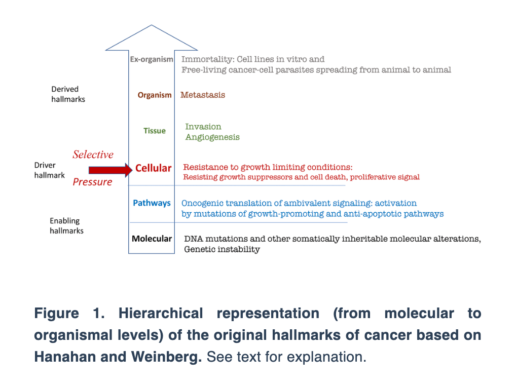
In this review, Dr. Blagosklonny expands on Gems and de Magalhães’ notion that canonic hallmarks of aging are superficial imitations of the hallmarks of cancer. He takes their work a step further and proposes the hallmarks of cancer and aging based on a hierarchical principle and the hyperfunction theory.
“Here I present the hallmarks of cancer, depicted as a circle by Hanahan and Weinberg [1], not as the circle but hierarchically, from molecular levels to the organism (Figure 1).”
Next, Dr. Blagosklonny depicts the hallmarks of aging suggested by López-Otín et al. based on the hierarchical principle.
“This representation renders hallmarks tangible but reveals three shortcomings (Figure 2).”
The first shortcoming that Dr. Blagosklonny notes is the lack of hallmarks on the organismal level. The second is that the relationship between hallmarks on different levels is unclear. The third is that the inclusion of genetic instability as a hallmark is based on the theory that aging is caused by the accumulation of molecular damage.
“The molecular damage theory was refuted by key experiments, as discussed in detail [44–51].”
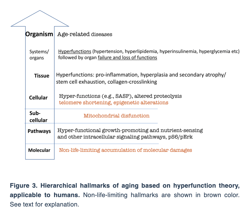
Dr. Blagosklonny then uses the hyperfunction theory to arrange the hierarchical hallmarks of aging.
“Let us depict hallmarks of aging, according to the hyperfunction theory of aging (Figure 3).”
Dr. Blagosklonny continues by discussing the key to understanding aging and aging as a selective force for cancer. He concludes this review by discussing the common hallmarks of cancer, aging and cell senescence.
“In organismal aging, cancer and cellular senescence, the same key signaling pathways, such as mTOR, are involved. This is why the same drugs, such as rapamycin, can suppress all of them.”
Launched in 2009, Aging-US publishes papers of general interest and biological significance in all fields of aging research and age-related diseases, including cancer—and now, with a special focus on COVID-19 vulnerability as an age-dependent syndrome. Topics in Aging-US go beyond traditional gerontology, including, but not limited to, cellular and molecular biology, human age-related diseases, pathology in model organisms, signal transduction pathways (e.g., p53, sirtuins, and PI-3K/AKT/mTOR, among others), and approaches to modulating these signaling pathways.
Published on the cover of Aging’s Volume 14, Issue 7, researchers conducted a new study investigating the role of IGFBP5 in replicative senescence.
The Trending With Impact series highlights Aging (Aging-US) publications that attract higher visibility among readers around the world online, in the news, and on social media—beyond normal readership levels. Look for future science news about the latest trending publications here, and at Aging-US.com.
—
Listen to an audio version of this article
In 1961, Leonard Hayflick and Paul Moorhead proposed a theory later named the Hayflick Limit. They discovered that a normal human cell can divide between 50 and 70 times before it can no longer proliferate and eventually dies. Researchers have since continued to explore this phenomenon and, today, this aging process is known as cellular (replicative) senescence.
“There are currently several experimental models of cellular senescence. Hayflick and Moorhead observed that primary human fibroblasts in culture exhibit a limited proliferative capacity [6]. This growth arrest during passages is called replicative senescence.”
This permanent cessation of the cell cycle is universally found in biology due to known and unknown causes, including the shortening of telomeres. While telomere shortening plays an important role, it is not the only event responsible for inducing cellular senescence. Thus, researchers have spent decades under the microscope experimenting with cellular models of replicative senescence.
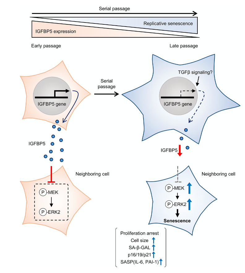
Cellular senescence is typically characterized by cell growth arrest, an increase of cells positive for SA-β -gal staining, and upregulation of p16 and p19. To begin this study, the team cultured embryonic mouse fibroblasts (MEFs) and conducted cell passages according to the 3T3 method. They found that the MEFs underwent senescence after the 5th passage (P5). The team also found that at P8, the expression of insulin-like growth factor binding protein 5 (IGFBP5) mRNA was significantly reduced when compared with that of P2 MEFs.
Next, the team performed a knockdown of IGFBP5 in the MEF cells. Results showed that IGFBP5 knockdown induced premature cellular senescence in P2 MEFs. Knockdown of IGFBP5 increased phosphorylation of extracellular signal-regulated kinases 1 and 2 (ERK1/2) but did not affect expression levels of Akt or p16 repressors. The researchers also found that supplementing the cell culture growth medium with additional exogenous IGFBP5 delayed growth arrest and reduced replicative senescence in the MEF cells.
“To examine whether activated ERK1 and ERK2 by IGFBP5 knockdown are involved in the induction of senescent phenotypes, we examined effects of knockdown of ERK1 and ERK2 using a combination with IGFBP5 siRNA in P2 MEFs.”
Upon further analysis of ERK1/2’s role in IGFBP5-knockdown cells, the team found that the silencing of ERK2, and not ERK1, blocked the increase in the number of SA-β-GAL-positive cells. ERK2 knockdown attenuated the reduction in the cell number and upregulation of p16 and p21 in IGFBP5-knockdown cells. This study provides evidence that downregulation of IGFBP5 contributes to replicative senescence via ERK2 activation in mouse embryonic fibroblasts.
Conclusion
For the first time, the role of IGFBP5 in replicative senescence was demonstrated in MEFs. Their findings suggest that ERK2 underlies cellular senescence induced by IGFBP5 downregulation. Cellular senescence appears to be a complex process with many moving parts. While more research is needed to fully understand the role of IGFBP5 in replicative senescence, this study provides new insights into the underlying mechanisms involved in this complex process.
“In conclusion, the results of the present study demonstrated that downregulation of IGFBP5 during serial passage contributes to replicative senescence via an ERK2-dependent mechanism (Figure 6). The results suggest that IGFBP5 counteracts replicative senescence in MEFs.”
Figure 6. Schematic summary of our findings. MEFs at early passage secrete certain levels of IGFBP5. Secreted IGFBP5 proteins inhibit MEK/ERK2 by attenuating their phosphorylation (P) in the neighboring cell, leading to suppression of cellular senescence. IGFBP5 secretion is decreased during serial passage, causing activation of ERK2 and cellular senescence.
Click here to read the full research paper published by Aging (Aging-US).
Aging (Aging-US) is an open-access journal that publishes research papers bi-monthly in all fields of aging research. These papers are available at no cost to readers on Aging-us.com. Open-access journals have the power to benefit humanity from the inside out by rapidly disseminating information that may be freely shared with researchers, colleagues, family, and friends around the world.
Researchers examined the effects of thoracic radiation-induced senescent cells on tumor progression, and the role of senotherapeutics to mitigate these effects.
Radiation therapy, advanced medical linear accelerator in therapeutic oncology to treat cancer
The Trending With Impact series highlights Aging (Aging-US) publications that attract higher visibility among readers around the world online, in the news, and on social media—beyond normal readership levels. Look for future science news about the latest trending publications here, and at Aging-US.com.
—
Listen to an audio version of this article
Radiation therapy is a highly-efficacious inducer of cancer cell death. With this being said, radiation has also previously been shown to cause premature senescence in the lung parenchyma. Senescence in cancer cells was previously only thought of as a mechanism capable of suppressing tumor cell proliferation by halting the cell cycle. However, a growing body of evidence shows that senescent cells may play a pro-tumorigenic role in cancer.
In the tumor microenvironment, the accumulation of senescent cells can become tumorigenic due to a lack of normal tissue stem cells and due to the expression of the senescence-associated secretory phenotype (SASP). SASP expression is when senescent cells secrete high levels of inflammatory cytokines, immune modulators, growth factors, and proteases. In addition to reinforcing senescence, SASP can create a biological environment that is immuno-suppressed and tumor-permissive. Radiation-induced senescence has previously been shown to have negative impacts on cancer patients.
“Cells that have undergone premature senescence due to stress, such as irradiation, are resistant to apoptotic cell death and effectively escape immune surveillance, resulting in their accumulation in tissue over time.”
Recently, researchers from the National Cancer Institute investigated the irradiated lung and the impact of radiation-induced senescent parenchymal cells on tumor growth. They also explored three senotherapeutics, rapamycin, INK-128 and ABT-737, for their potential to mitigate radiation-induced senescence. On February 12, 2022, the team’s priority research paper was published on the cover of Aging (Aging-US)Volume 14, Issue 3, and entitled, “Senescence-associated tumor growth is promoted by 12-Lipoxygenase.”
The Study
In this study, researchers intravenously injected melanoma cells into murine models two, four and eight weeks after daily fractions of thoracic irradiation exposure. There was also a control arm of unirradiated murine models. Tumor development was monitored by the number and size of the nodules in lung tissues. The number of cells exhibiting senescent activity was also recorded after two, four and eight weeks of thoracic irradiation. Their data demonstrated a correlation between the time points when tumors developed in the irradiated lungs and a marked accumulation of senescent cells.
“As previously described, in irradiated lungs, senescent cells increased significantly 4 and 8 weeks after IR compared to age matched unirradiated controls (Figure 1A).”
A characteristic of oncogene- and stress-induced senescence is the activation of mTOR signaling. Given this connection, the researchers conducted parallel studies evaluating senostatic agents capable of targeting the mTOR pathway, rapamycin and INK-128, and a senolytic agent to selectively eliminate senescent cells, ABT-737. The data showed that rapamycin and INK-128 significantly reduced the number of tumor nodules in the lungs of irradiated mice compared to the controls. ABT-737 demonstrated reduced pulmonary senescence in irradiated mice.
The researchers also studied 12-Lipoxygensae (12-LOX), an enzyme that metabolizes a certain SASP molecule previously implicated in pulmonary senescence: 12(S)-HETE. 12-LOX is a known contributor to radiation-induced senescence and lung injury. The team specifically focused on the role of 12-LOX in pulmonary senescence and its impact on radiation-enhanced tumor growth. They found that inhibiting 12-LOX activity reduced radiation-induced lung senescence and mitigated radiation-enhanced tumor growth.
“Finally, we link senescence associated 12-LOX activity and production of 12(S)-HETE to the observed enhanced tumor growth after irradiation.”
Conclusion
In sum, the researchers found that radiation therapy can induce senescence in the lung parenchyma and also enhance tumor growth. The contribution of senescence in tumor progression was emphasized by the protection delivered by the mTOR-targeted senostatic and senolytic agents. This important discovery could lead to new therapies for cancer patients who are undergoing radiation therapy.
“Together, this study demonstrates the critical role of senescence in mediating radiation-enhanced tumor growth and identifies Alox12 as an important player in this phenomenon. Treatment with a senostatic agent, INK-128, identified in this study, or with agents like rapamycin and ABT-737 suggested their potential therapeutic use in alleviating radiation associated tumor growth.”
Click here to read the full priority research paper published by Aging (Aging-US).
Aging (Aging-US) is an open-access journal that publishes research papers bi-monthly in all fields of aging research. These papers are available to read at no cost to readers on Aging-us.com. Open-access journals offer information that has the potential to benefit our societies from the inside out and may be shared with friends, neighbors, colleagues, and other researchers, far and wide.