Aging-US: Hallmarks of Cancer and Hallmarks of Aging

“Hyperfunctional signaling directly drives age-related diseases.”

— Mikhail Blagosklonny, M.D., Ph.D.

Listen to an audio version of this press release

BUFFALO, NY- May 18, 2022 – Dr. Mikhail Blagosklonny published his new review paper in Aging (Aging-US) Volume 14, Issue 9, entitled, “Hallmarks of cancer and hallmarks of aging.”

In this review, Dr. Blagosklonny expands on Gems and de Magalhães’ notion that canonic hallmarks of aging are superficial imitations of the hallmarks of cancer. He takes their work a step further and proposes the hallmarks of cancer and aging based on a hierarchical principle and the hyperfunction theory.

“Here I present the hallmarks of cancer, depicted as a circle by Hanahan and Weinberg [1], not as the circle but hierarchically, from molecular levels to the organism (Figure 1).”

Figure 1. Hierarchical representation (from molecular to organismal levels) of the original hallmarks of cancer based on Hanahan and Weinberg. See text for explanation.

Next, Dr. Blagosklonny depicts the hallmarks of aging suggested by López-Otín et al. based on the hierarchical principle. 

“This representation renders hallmarks tangible but reveals three shortcomings (Figure 2).”

Figure 2. Hierarchical representation of the hallmarks of aging based on López-Otín et al. See text for explanation.

The first shortcoming that Dr. Blagosklonny notes is the lack of hallmarks on the organismal level. The second is that the relationship between hallmarks on different levels is unclear. The third is that the inclusion of genetic instability as a hallmark is based on the theory that aging is caused by the accumulation of molecular damage. 

“The molecular damage theory was refuted by key experiments, as discussed in detail [44–51].” 

Dr. Blagosklonny then uses the hyperfunction theory to arrange the hierarchical hallmarks of aging.

“Let us depict hallmarks of aging, according to the hyperfunction theory of aging (Figure 3).”

Figure 3. Hierarchical hallmarks of aging based on hyperfunction theory, applicable to humans. Non-life-limiting hallmarks are shown in brown color. See text for explanation.

Dr. Blagosklonny continues by discussing the key to understanding aging and aging as a selective force for cancer. He concludes this review by discussing the common hallmarks of cancer, aging and cell senescence.

“In organismal aging, cancer and cellular senescence, the same key signaling pathways, such as mTOR, are involved. This is why the same drugs, such as rapamycin, can suppress all of them.”

DOI: https://doi.org/10.18632/aging.204082 

Correspondence to: Mikhail V. Blagosklonny 

Email: Blagosklonny@oncotarget.comBlagosklonny@rapalogs.com 

Keywords: oncology, carcinogenesis, geroscience, mTOR, rapamycin, hyperfunction theory

Follow Dr. Blagosklonny on Twitter: https://twitter.com/Blagosklonny

AGING (AGING-US) VIDEOS: YouTube | LabTube | Aging-US.com

About Aging-US:

Launched in 2009, Aging-US publishes papers of general interest and biological significance in all fields of aging research and age-related diseases, including cancer—and now, with a special focus on COVID-19 vulnerability as an age-dependent syndrome. Topics in Aging-US go beyond traditional gerontology, including, but not limited to, cellular and molecular biology, human age-related diseases, pathology in model organisms, signal transduction pathways (e.g., p53, sirtuins, and PI-3K/AKT/mTOR, among others), and approaches to modulating these signaling pathways.

Follow Aging on social media: 

For media inquiries, please contact media@impactjournals.com.

Aging (Aging-US) Journal Office

6666 E. Quaker Str., Suite 1B

Orchard Park, NY 14127

Phone: 1-800-922-0957, option 1
###

TP53 Restoration Sensitizes Pancreatic Cancer to Multiple Drugs

Listen to an audio version of this article

Patients over the age of 50 years old who have been diagnosed with pancreatic cancer have a poorer rate of survival compared to younger patients. This means that pancreatic cancer is a disease associated with aging. The most common type of pancreatic cancer is pancreatic ductal adenocarcinoma (PDAC) and it is frequently diagnosed in its later stages. PDAC is often refractive to chemotherapies and develops resistance to inhibitors and other drugs. Therefore, there is a critical need for researchers to discover novel strategies to overcome drug resistance in PDAC cells.

One potential strategy is to focus on a key gene known for its involvement in many cell processes, including drug resistance and metabolism: TP53. The TP53 gene is often mutated or deleted in cancer cells, which can lead to drug resistance and cancer metastasis. In PDACS, this tumor suppressor gene has been shown to be mutated in 50–75% of patients.

“Many genes have been implicated in PDAC including KRAS, TP53, CDKN2A, SMAD4 and PDGFβR [3, 8, 9, 1822].”

In a new study, researchers—from Brody School of Medicine at East Carolina University, Università di Bologna, University of Parma, and University of Wroclaw—further elucidated TP53’s role in drug resistance in PDAC cells. On April 27, 2022, their research paper was published in Aging (Aging-US) on the cover of Volume 14, Issue 8, and entitled, “Wild type and gain of function mutant TP53 can regulate the sensitivity of pancreatic cancer cells to chemotherapeutic drugs, EGFR/Ras/Raf/MEK, and PI3K/mTORC1/GSK-3 pathway inhibitors, nutraceuticals and alter metabolic properties.”

The Study

In these in vitro studies, the researchers cultured two different PDAC cell lines. One cell line had a gain of function (GOF) TP53 mutation (MIA-PaCa-2) and the other had a loss of TP53 (PANC-28). Both PDAC cell lines also have activating mutations in the KRAS gene. Next, the team introduced either wild-type TP53 (WT-TP53) or a control vector into both PDAC cell lines. Effects from this experiment were analyzed using 26 clinically approved agents.

The chemotherapeutic drugs included: Docetaxel, 5-fluorouracil (5-FU), gemcitabine, Aclacinomycin, Doxorubicin, and Cisplatin. The signal transduction inhibitors included: ARS-1620, PD0325901, LY294002, Pifithrin-μ, 6-bromoindirubin-30-oxime (BIO), SB415286, CHIR99021, Rapamycin, AG1498, Gilteritinib, Sorafenib, OTX008, Tiplaxtinin, Verapamil, and Vismodegib. The natural products included: Cyclopamine, Parthenolide2, Isoliquiritin2, Genistein2, and Daidzein2. The researchers also illustrated the effects of WT-TP53 and mutant TP53 on PDAC cell metabolism with metformin and rapamycin.

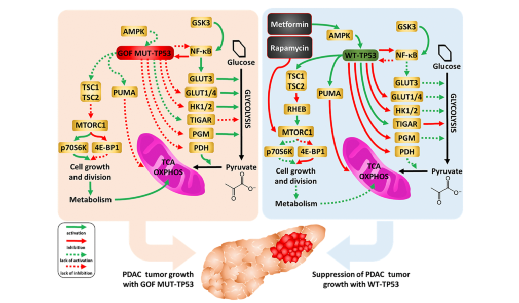
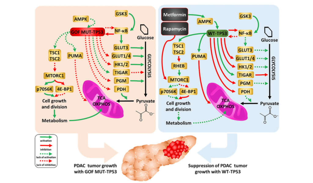
“An overview of the effects of WT and mutant TP53 on metabolic properties, together with the effects of metformin and rapamycin, and drugs used to inhibit pancreatic cancer growth, is presented in Figure 16.”

Figure 16. Influences of mutant and WT-TP53 on mitochondrial activity and glucose metabolism and effects of rapamycin and metformin. The effects of WT and mutant TP53 on key enzymes important in glycolysis and how they can influence metabolism and PDAC tumor growth. In our studies, we have examined the effect of GOF mutant TP53 and in some cases WT TP53. In addition, sites of interaction of the type 2 diabetes drug metformin and the immunosuppressive drug rapamycin and their effects on AMPK and mTORC1 are indicated. TP53 can induce mitochondrial apoptosis pathway by regulating the expression of PUMA and other proteins.

The Results

The researchers found that, in the presence of chemotherapeutic drugs, PDAC clonogenicity was decreased by the restoration of WT-TP53. Overall, the restoration of WT-TP53 in PDAC cells increased sensitivity/decreased resistance to various chemotherapeutic drugs, inhibitors and natural products. WT-TP53 also influenced  PDAC cell metabolic properties, including their metabolism. The authors also noted that the activity of mTORC1 (target of rapamycin), which is important in cellular growth and metabolism, can be affected by mutant TP53. They found that GOF mutated TP53 may render PDAC cells more resistant to rapamycin.

“Rapamycin and metformin can interfere with some of the important pathways in the mitochondria, some of which are regulated by TP53 [9698].”

Conclusion

Overall, these results suggest that WT-TP53 can play a key role in PDAC cell sensitivity to multiple drugs used to treat pancreatic cancer. Further studies are needed to better understand the mechanisms underlying the effects of TP53 on drug resistance and metabolism in PDAC cells, as well as its clinical implications.

“Regardless of which of the above processes contributes more to the reduction of mitochondrial metabolism in comparison with the same cells that only express GOF TP53, together the observed changes suggest restoration of WT-TP3 activity confers increased sensitization to various drugs and therapeutic molecules, natural products as well as nutraceuticals.”

Click here to read the full research paper published by Aging (Aging-US).

AGING (AGING-US) VIDEOS: YouTube | LabTube | Aging-US.com

Aging (Aging-US) is an open-access journal that publishes research papers bi-monthly in all fields of aging research. These papers are available at no cost to readers on Aging-us.com. Open-access journals have the power to benefit humanity from the inside out by rapidly disseminating information that may be freely shared with researchers, colleagues, family, and friends around the world.

For media inquiries, please contact media@impactjournals.com.

Trending With Impact: Radiation, Senescence and Senotherapeutics

Researchers examined the effects of thoracic radiation-induced senescent cells on tumor progression, and the role of senotherapeutics to mitigate these effects.

Radiation therapy, advanced medical linear accelerator in therapeutic oncology to treat cancer

The Trending With Impact series highlights Aging (Aging-US) publications that attract higher visibility among readers around the world online, in the news, and on social media—beyond normal readership levels. Look for future science news about the latest trending publications here, and at Aging-US.com.

Listen to an audio version of this article

Radiation therapy is a highly-efficacious inducer of cancer cell death. With this being said, radiation has also previously been shown to cause premature senescence in the lung parenchyma. Senescence in cancer cells was previously only thought of as a mechanism capable of suppressing tumor cell proliferation by halting the cell cycle. However, a growing body of evidence shows that senescent cells may play a pro-tumorigenic role in cancer.

In the tumor microenvironment, the accumulation of senescent cells can become tumorigenic due to a lack of normal tissue stem cells and due to the expression of the senescence-associated secretory phenotype (SASP). SASP expression is when senescent cells secrete high levels of inflammatory cytokines, immune modulators, growth factors, and proteases. In addition to reinforcing senescence, SASP can create a biological environment that is immuno-suppressed and tumor-permissive. Radiation-induced senescence has previously been shown to have negative impacts on cancer patients.

“Cells that have undergone premature senescence due to stress, such as irradiation, are resistant to apoptotic cell death and effectively escape immune surveillance, resulting in their accumulation in tissue over time.”

Recently, researchers from the National Cancer Institute investigated the irradiated lung and the impact of radiation-induced senescent parenchymal cells on tumor growth. They also explored three senotherapeutics, rapamycin, INK-128 and ABT-737, for their potential to mitigate radiation-induced senescence. On February 12, 2022, the team’s priority research paper was published on the cover of Aging (Aging-US) Volume 14, Issue 3, and entitled, “Senescence-associated tumor growth is promoted by 12-Lipoxygenase.”

The Study

In this study, researchers intravenously injected melanoma cells into murine models two, four and eight weeks after daily fractions of thoracic irradiation exposure. There was also a control arm of unirradiated murine models. Tumor development was monitored by the number and size of the nodules in lung tissues. The number of cells exhibiting senescent activity was also recorded after two, four and eight weeks of thoracic irradiation. Their data demonstrated a correlation between the time points when tumors developed in the irradiated lungs and a marked accumulation of senescent cells.

“As previously described, in irradiated lungs, senescent cells increased significantly 4 and 8 weeks after IR compared to age matched unirradiated controls (Figure 1A).”

A characteristic of oncogene- and stress-induced senescence is the activation of mTOR signaling. Given this connection, the researchers conducted parallel studies evaluating senostatic agents capable of targeting the mTOR pathway, rapamycin and INK-128, and a senolytic agent to selectively eliminate senescent cells, ABT-737.  The data showed that rapamycin and INK-128 significantly reduced the number of tumor nodules in the lungs of irradiated mice compared to the controls. ABT-737 demonstrated reduced pulmonary senescence in irradiated mice.

The researchers also studied 12-Lipoxygensae (12-LOX), an enzyme that metabolizes a certain SASP molecule previously implicated in pulmonary senescence: 12(S)-HETE. 12-LOX is a known contributor to radiation-induced senescence and lung injury. The team specifically focused on the role of 12-LOX in pulmonary senescence and its impact on radiation-enhanced tumor growth. They found that inhibiting 12-LOX activity reduced radiation-induced lung senescence and mitigated radiation-enhanced tumor growth.

“Finally, we link senescence associated 12-LOX activity and production of 12(S)-HETE to the observed enhanced tumor growth after irradiation.”

Conclusion

In sum, the researchers found that radiation therapy can induce senescence in the lung parenchyma and also enhance tumor growth. The contribution of senescence in tumor progression was emphasized by the protection delivered by the mTOR-targeted senostatic and senolytic agents. This important discovery could lead to new therapies for cancer patients who are undergoing radiation therapy.

“Together, this study demonstrates the critical role of senescence in mediating radiation-enhanced tumor growth and identifies Alox12 as an important player in this phenomenon. Treatment with a senostatic agent, INK-128, identified in this study, or with agents like rapamycin and ABT-737 suggested their potential therapeutic use in alleviating radiation associated tumor growth.”

Click here to read the full priority research paper published by Aging (Aging-US).

AGING (AGING-US) VIDEOS: YouTube | LabTube | Aging-US.com

Aging (Aging-US) is an open-access journal that publishes research papers bi-monthly in all fields of aging research. These papers are available to read at no cost to readers on Aging-us.com. Open-access journals offer information that has the potential to benefit our societies from the inside out and may be shared with friends, neighbors, colleagues, and other researchers, far and wide.

For media inquiries, please contact media@impactjournals.com.

Rapamycin Rules out DNA Damage Theory of Aging

Dr. Mikhail Blagosklonny gleans an important new discovery in aging research—deduced from recent studies on short-lived mice and rapamycin.

3D illustration of a mutated or damaged DNA strand

The Top-Performer series highlights papers published by Aging that have generated a high Altmetric attention score. Altmetric scores, located at the top-left of trending Aging papers, provide an at-a-glance indication of the volume and type of online attention the research has received.

Read Aging’s Top 100 Altmetric papers.

Listen to an audio version of this article

The exact mechanisms at play in the human aging process are still up for debate. A number of great minds in science have proposed plausible aging mechanisms and theories, such as DNA damage, telomere shortening, and DNA damage theories of aging. DNA damage theories suggest that aging is functional decline, caused by the accumulation of molecular damage. However, some scientists counterclaim that neither DNA damage nor telomere shortening limit lifespan or cause aging.

Dr. Mikhail Blagosklonny—an adjunct faculty member at the Roswell Park Comprehensive Cancer Center and Editor-in-Chief at Aging, Oncotarget, Oncoscience, and Cell Cycle—gleaned an important new perspective from recent aging studies, which could have been overlooked. He expanded on this discovery in a recent research perspective that was published in February 2021 in an issue of Aging, entitled: “DNA- and telomere-damage does not limit lifespan: evidence from rapamycin.” To date, this research paper has generated an Altmetric Attention score of 43.

Rapamycin is a macrolide antibiotic that has immunosuppressive properties, regulates a key cellular growth pathway (mTOR), and has been at the center of numerous studies of aging since its discovery in 1964. Dr. Blagosklonny explains that, based on findings from recent mouse-model studies of rapamycin’s effects on short-lived mice, normal aging is not caused by the accumulation of molecular damage or telomere shortening.

“Here I discussed new evidence that normal aging is not caused by accumulation of molecular damage or telomere shortening: while extending normal lifespan in mice, rapamycin failed to do so in mice dying from molecular damage (Figure 1).”

Evidence From Rapamycin

In the study which Dr. Blagosklonny refers to, researchers genetically modified mice to artificially shorten telomeres, administered rapamycin to normal mice and the telomerase-deficient short-lived mice, and observed the effects. In normal mice, results were congruent with a number of other studies that found lifespan was significantly extended. In the telomerase-deficient mice, lifespan was shortened as a result of rapamycin. 

“While shortening lifespan by 18% in unnatural telomerase-deficient mice, in the same study in natural mice, rapamycin increased lifespan by 39% and healthspan by 58% (measured as tumor-free survival) [3].”

Given that rapamycin prolongs life in normal mice, Dr. Blagosklonny asserts that this study proves that normal lifespan is not constrained by telomere length. Telomeres only become life-limiting when they are artificially shortened to the point where rapamycin can no longer extend lifespan. Furthermore, Dr. Blagosklonny explains that although molecular damage and telomere shortening could be life-limiting, they ultimately do not limit life because quasi-programmed aging occurs at a faster rate.

“Although molecular damage accumulates, this accumulation is not life-limiting because quasi-programmed aging terminates life first (Figure 1A). Quasi-programmed (hyperfunctional) aging is life-limiting, because it is favored by natural selection.”

Quasi-Programmed (Hyperfunctional) Aging

In 2012, Dr. Blagosklonny wrote another widely-read research perspective that explains in great detail what his proposed hyperfunction theory of aging is, entitled, “Answering the ultimate question “What is the Proximal Cause of Aging?

“According to hyperfunction theory, aging is quasi-programmed, a continuation of developmental growth programs, driven in part by hyper-functional signaling pathways including the mTOR pathway [9].”

He explains that hyperfunction is an excessive, yet normal function that occurs later in life. Hyperfunction in this context does not necessarily mean an increase in function and, in some cases, it even means a decrease in function. The same pathways and functions that drive growth and development earlier in life, also drive age-related diseases later in life. Dr. Blagosklonny proposes that quasi-programmed (hyperfunctional) aging is favored by natural selection and is what limits life.

“It is hyperfunctional signaling pathways such as mTOR (one of many) that drive both growth and aging, causing age-related diseases that in turn damage organs, leading to secondary loss of function.”

Many signaling pathways interact with mTOR to drive aging, forming a network, including MEK/MAPK, NF-kB, p63, HIF-1, and many others. Dr. Blagosklonny suggests that, in theory, there could be a number of mTOR-independent factors of quasi-programmed aging that are life-limiting, as well. He goes on to exemplify several lines of evidence concluding that it is not molecular damage that causes normal aging or limits life—it is normal, quasi-programmed (hyperfunctional) aging.

Conclusion

Dr. Blagosklonny mentions a forthcoming review that will be entitled: “When longevity drugs do not increase longevity: Unifying development-driven and damage-induced theories of aging.”

“Once again, damage accumulates and must cause death eventually, but quasi-programmed (hyperfunctional) aging terminates life first. Molecular damage can become life-limiting, when artificially accelerated or, potentially, when quasi-programmed aging is decelerated.” 

Click here to read the full research perspective, published in Aging.

Aging is an open-access journal that publishes research papers monthly in all fields of aging research and other topics. These papers are available to read at no cost to readers on Aging-us.com. Open-access journals offer information that has the potential to benefit our societies from the inside out and may be shared with friends, neighbors, colleagues, and other researchers, far and wide.

For media inquiries, please contact media@impactjournals.com.

Aging is a proud participant in the AACR Annual Meeting 2021 #AACR21
  • Follow Us