Predicting Brain Age With Machine Learning and Transcriptome Profiling

In this study, researchers investigated age-associated gene expression changes in the prefrontal cortex of male and female brains and used machine learning to develop age prediction models.

The human brain is a complex organ, and its aging process is influenced by a plethora of factors, both genetic and environmental. Aging-related changes in the brain can lead to cognitive decline and susceptibility to neurodegenerative diseases. Therefore, understanding the molecular mechanisms underlying these changes is crucial for developing therapeutic strategies to delay or prevent age-related cognitive decline.

Over the past few years, a myriad of scientific studies have been conducted to understand the intricate relationship between our genes and the aging process. In a new study, researchers Joseph A. Zarrella and Amy Tsurumi from Harvard T.H. Chan School of Public Health, Massachusetts General Hospital, Harvard Medical School, and Shriner’s Hospitals for Children-Boston explored the concept of genome brain age prediction, a groundbreaking area of study that employs advanced bioinformatics tools to analyze changes in gene expression associated with aging. On February 28, 2024, their research paper was published and chosen as the cover paper for Aging’s Volume 16, Issue 5, entitled, “Genome-wide transcriptome profiling and development of age prediction models in the human brain.”

“[…] we aimed to profile transcriptome changes in the aging PFC [prefrontal cortex] overall and compare females and males, and develop prediction models for age.”

Transcriptome Profiling in the Prefrontal Cortex

The prefrontal cortex (PFC) plays a significant role in the aging process. It is responsible for a host of cognitive functions, including decision-making and planning. Throughout the aging process, significant transcriptome alterations occur in the PFC compared to other regions of the brain. These alterations can influence cognitive decline and susceptibility to neurodegenerative diseases.

Delving deeper into the complexities of aging, researchers have turned to transcriptome profiling as a powerful tool to uncover the molecular changes occurring within the prefrontal cortex. Transcriptome profiling allows scientists to measure the expression levels of all genes in a cell or tissue. By analyzing the transcriptome of the PFC, researchers can identify genes that are differentially expressed during the aging process. These genes can serve as potential biomarkers for age prediction.

The Study

In their groundbreaking research, Zarrella and Tsurumi aimed to develop prediction models for age based on the expression levels of specific panels of transcripts in the PFC. They leveraged advanced machine learning algorithms, including the least absolute shrinkage and selection operator (Lasso), Elastic Net (EN), eXtreme Gradient Boosting (XGBoost), and Light Gradient Boosting Machine (LightGBM), to develop accurate prediction models for chronological age.

The researchers used postmortem PFC transcriptome datasets obtained from the Gene Expression Omnibus (GEO) repository, ranging in age from 21 to 105 years. They identified differentially regulated transcripts in old and elderly samples compared to young samples and assessed the genes associated with age using ontology, pathway, and network analyses.

Machine learning algorithms were used to develop accurate prediction models for chronological age based on the expression levels of specific transcripts. The study found that specific gene expression changes in the PFC are highly correlated with age. Some transcripts showed female and male-specific differences, indicating that sex may play a role in the aging process at the molecular level.

Key Findings & Implications

The study identified several key genes whose expression levels change significantly with age. These genes include Carbonic Anhydrase 4 (CA4), Calbindin 1 (CALB1), Neuropilin and Tolloid Like 2 (NETO2), and Olfactomedin1 (OLFM1), among others. Many of these genes have been previously implicated in aging or aging-related diseases, validating the results of this study.

The researchers also developed four highly accurate age prediction models using different machine learning algorithms. These models were validated in a test set and an external validation set, demonstrating their potential application in predicting chronological age based on gene expression levels.

“Our results support the notions that specific gene expression changes in the PFC are highly correlated with age, that some transcripts show female and male-specific differences, and that machine learning algorithms are useful tools for developing prediction models for age based on transcriptome information.”

Conclusions & Future Directions

This study sheds light on the complex relationship between gene expression changes and the aging process in the human brain. The findings underscore the potential of using transcriptome profiling and machine learning algorithms for age prediction. The identified genes could serve as potential biomarkers for age prediction and may offer new insights into the molecular mechanisms underlying the aging process.

However, further validation of these models in larger populations and molecular studies to elucidate the potential mechanisms by which the identified transcripts may be related to aging phenotypes would be beneficial. Additionally, more inclusive studies investigating the interplay between genetic markers and factors such as sex, lifestyle, and environmental exposures are warranted.

In conclusion, this study provides a promising foundation for future research on genome brain age prediction. It also underscores the potential of transcriptome profiling and machine learning for exploring the complex interplay between our genes and the aging process. This approach could pave the way for personalized medicine strategies aimed at preventing or delaying age-related cognitive decline and neurodegenerative diseases.

Click here to read the full research paper published in Aging.

Aging is an open-access, traditional, peer-reviewed journal that publishes high-impact papers in all fields of aging research. All papers are available to readers (at no cost and free of subscription barriers) in bi-monthly issues at Aging-US.com.

Click here to subscribe to Aging publication updates.

For media inquiries, please contact media@impactjournals.com.

Brain Aging Insights from Individuals Without Neurodegeneration

The Trending With Impact series highlights Aging publications (listed by MEDLINE/PubMed as “Aging (Albany NY)” and “Aging-US” by Web of Science) that attract higher visibility among readers around the world online, in the news and on social media—beyond normal readership levels. Look for future science news about the latest trending publications here, and at Aging-US.com.

Listen to an audio version of this article

A healthy brain continuously produces new proteins to support synaptic plasticity, maintain neuronal health, facilitate signaling pathways, produce neurotransmitters, enable neuroplasticity and adaptation, and meet its metabolic demands. These processes are essential for normal brain function, learning, memory, and overall cognitive abilities. Researchers believe that the dysregulation of proteins is at the core of brain aging. However, the exact recipe for protein dysregulation that leads to accelerated brain aging and neurodegenerative disorders has yet to be brought to light. 

Previous brain proteostasis (referring to the maintenance of protein homeostasis in brain cells) studies in individuals with Alzheimer’s disease (AD) pathology and age-related neuropathological changes have shown protein dysregulation leading to a buildup of amyloid plaques and neurofibrillary tangles. While these studies have greatly enhanced our knowledge of brain aging, gaps in our understanding remain. What proteomic characteristics do healthy brain aging individuals—without neurodegenerative disorders—have in common?

“To our knowledge, whole phosphoproteomes centered on the human brain aging without AD pathology are unavailable.”

In a new study, researchers Pol Andrés-Benito, Ignacio Íñigo-Marco, Marta Brullas, Margarita Carmona, José Antonio del Rio, Joaquín Fernández-Irigoyen, Enrique Santamaría, Mónica Povedano, and Isidro Ferrer from Bellvitge Institute for Biomedical Research, Universidad Pública de Navarra, Barcelona Institute for Science and Technology, and University of Barcelona aimed to shed light on the mechanisms underlying brain aging in the absence of AD pathology and age-related neuropathological changes. Their research paper was published on May 13, 2023, in Aging’s Volume 15, Issue 9, and entitled, “Proteostatic modulation in brain aging without associated Alzheimer’s disease-and age-related neuropathological changes.”

The Study

The production of new proteins is crucial for maintaining protein homeostasis in the brain. A post-translational modification used to maintain this homeostasis is protein phosphorylation. In this study, the researchers conducted proteomic and phosphoproteomic analyses of frontal cortex samples from the donor brains of deceased individuals between the ages of 30 and 85. These individuals had passed away due to non-neurological complications and were reported to have had full cognitive function. Individuals were divided into four groups: young group one (30–44), middle-aged group two (45-52), early-elderly group three (64–70), and late-elderly group four (75–85).

“​​We chose the FC [frontal cortex] because of its role in cognition and emotion and the abundant molecular information that permits comparison with other studies.”

Conventional label-free- and SWATH- (sequential window acquisition of all theoretical fragment ion spectra) mass spectrometry were used to assess the (phospho)proteomes of the frontal cortices from individuals in all four age groups. Immunohistochemistry and/or western blotting was/were also used to validate a subgroup of proteins. The researchers categorized deregulated proteins and phosphoproteins into eight clusters based on their age-dependent expression similarity (see paper for clusters). Interestingly, protein and phosphoprotein levels of the larger hierarchical clusters were stable until the age of 70 years. After 70, the late-elderly group showed significant decreased or increased expression of protein clusters one and seven, and major phosphorylation modifications occurred in clusters four and eight.

Results

The team then used multi-comparative analyses to categorize altered proteins and phosphoproteins as neuronal, astroglial, oligodendroglial, microglial, and endothelial. They observed a similar pattern among proteomic and phosphoproteomic alterations: major changes were related to neuronal cell populations across all four groups—and these changes were more pronounced with age. Cytoskeletal and membrane proteins accounted for the largest number of differentially-expressed proteins and phosphoproteins.

“Furthermore, main alterations in the proteome are associated with proteins specific to neuronal populations, rather than those found in other cell types in the brain.”

Their findings also revealed a decline in the expression of P20S α + β with aging, while the expression of P19S and immunoproteasome subunits LMP2 and LMP7 remained preserved. Notably, the expression levels of an autophagy component, ATG5, remained unchanged with age. Some mitochondrial membrane proteins showed altered levels at advanced ages, but key markers of mitochondrial function were preserved. These findings suggest a potential preservation of these pathways in advanced aging, contrasting with observations in neurodegenerative disorders. Additionally, reduced levels of GSK3α/β were observed, and the researchers point out that this decrease in GSK3α/β with age may be understood as protective against different age-related brain diseases.

Summary & Conclusion

“Therefore, our results fill the gap between brain ageing without ADNC [AD neuropathological changes], and cases with early and advanced stages of AD pathology.”

The researchers are forthcoming about limitations of this study. Given it is rare for old-aged individuals not to have neurological deficits, AD or other neuropathological changes, their main limitation was that each of the four groups included merely four individuals. Despite limitations, these findings contribute to our understanding of brain aging in the absence of AD pathology and age-related neuropathological changes. 

The study revealed that major changes in protein expression were primarily associated with neuronal cell populations and became more pronounced with age. The preservation of specific protein pathways, proteasome components, autophagy-related components, and mitochondrial markers in advanced aging individuals without neurodegenerative disorders suggests the presence of resilience mechanisms that protect against protein dysregulation and neurodegeneration. Overall, this research provides valuable insights into the proteomic characteristics of healthy brain aging and highlights potential targets for therapeutic interventions aimed at promoting healthy brain aging and preventing age-related neurodegenerative diseases. Further studies are necessary to elucidate the specific mechanisms underlying these proteomic alterations and their functional implications in brain aging.

“The present observations identify proteostatic changes, including different changes in the phosphoproteome in the human FC in brain aging in the rare subpopulation of old-aged individuals without neurological deficits, and not having ADNC and other neuropathological change in any region of the telencephalon.”

Click here to read the full research paper published by Aging.

Aging is an open-access, peer-reviewed journal that has been publishing high-impact papers in all fields of aging research since 2009. These papers are available to readers (at no cost and free of subscription barriers) in bi-monthly issues at Aging-US.com.

Click here to subscribe to Aging publication updates.

For media inquiries, please contact media@impactjournals.com.

The Brain Age Gap

The Trending With Impact series highlights Aging publications (listed by MEDLINE/PubMed as “Aging (Albany NY)” and “Aging-US” by Web of Science) that attract higher visibility among readers around the world online, in the news and on social media—beyond normal readership levels. Look for future science news about the latest trending publications here, and at Aging-US.com.

Listen to an audio version of this article

Aging is a risk factor for many diseases, including Alzheimer’s disease (AD). While scientists have made some progress in understanding the physiology of aging and its relationship to AD and related disorders, our understanding remains incomplete (to say the least). It is possible that civilization is currently in the midst of an artificial intelligence (AI) and machine learning (ML) “boom.” Researchers are now using AI and ML technologies to elevate our comprehension of aging and aging-related diseases.

“Artificial intelligence (AI) and machine learning (ML) technologies can help us better understand these diseases and aging itself by using biological data from the brain or other sources to create a mapping between age and biological data.”

In a new editorial paper, researchers Jeyeon Lee, Leland R. Barnard and David T. Jones from the Mayo Clinic in Rochester, Minnesota, discuss a recent study they conducted and explore the potential of AI to revolutionize the field of geriatrics. Their editorial was published in Aging’s Volume 15, Issue 8, on April 3, 2023, entitled, “Artificial intelligence and the aging mind.”

Their Study

In a recent 2022 study, Lee, Barnard, Jones, and the rest of their team developed convolutional neural network-based brain age prediction models using a large collection of data from brain magnetic resonance imaging (MRI) and brain fluorodeoxyglucose positron-emission tomography (FDG-PET) in people aged from 26 to 98 years old. In a sample of cognitively normal individuals, the AI models showed accurate brain age estimation of which a mean absolute error (MAE; unit, years) was 3.08±0.14 for the FDG-based model and 3.49±0.16 for the MRI-based model. 

The team found that higher brain age gaps (the difference between biological age and chronological age) were estimated in cohorts with neurodegenerative disorders—including mild cognitive impairment (MCI), AD, frontotemporal dementia (FTD), and dementia with Lewy bodies (DLB)—than normal controls. The brain age gap was strongly associated with pathologic tau protein levels and cognitive test scores. This gap also showed longitudinal predictive ability for cognitive decline in AD-related disorders.

“Interestingly, the brain imaging patterns generating brain age gaps in AD showed higher similarity with normal aging than other neurodegenerative syndromes implying that AD might be more like an accelerated representation of biological aging than others.”

Summary & Conclusions

The study conducted by Lee, Barnard, Jones, and their team using neural network-based brain age prediction models has shown promising results in accurately estimating brain age and identifying differences between normal aging and neurodegenerative disorders. However, the authors of this editorial note that variations in data make creating a uniform language used to compare and contrast large sums of data very difficult.

“Although more research and optimization are needed to determine its clinical usefulness, the study of brain age has great potential as a tool for understanding brain aging and age-related diseases.”

In conclusion, aging is a complex process that increases the risk of Alzheimer’s disease and various diseases. Recent advancements in artificial intelligence and machine learning technologies offer new opportunities to better understand the underlying mechanisms of aging and aging-related disorders. This research opens up exciting possibilities for the future of geriatric care and improving the lives of aging populations. As technology continues to advance, it is likely that we will gain further insights into aging through the brain age gap, ultimately leading to better prevention, diagnosis and treatment options.

“The fact that the brain age gap is a comprehensive and intuitive measure of disease severity using biological data that is already being acquired in clinical practice, makes it an attractive biomarker for further development for clinical use [8].”

Click here to read the full editorial paper published by Aging.

Aging is an open-access, peer-reviewed journal that has been publishing high-impact papers in all fields of aging research since 2009. These papers are available to readers (at no cost and free of subscription barriers) in bi-monthly issues at Aging-US.com.

Click here to subscribe to Aging publication updates.

For media inquiries, please contact media@impactjournals.com.

High School Students Use AI to Make Aging and Glioblastoma Discoveries

In a breakthrough study, three high school students and Insilico researchers used generative artificial intelligence (AI) to help identify new therapeutic targets for glioblastoma multiforme (GBM) and aging.

High School Students Use AI to Make Aging and Glioblastoma Discoveries
Listen to an audio version of this article

Glioblastoma multiforme (GBM) is one of the most aggressive and fatal malignant brain tumors. With a median survival time of 15 months, only about 25% of patients survive for one year and less than 5% survive for five years. As people get older, the risk of developing GBM increases. The discovery of new drug targets for GBM is of paramount importance.

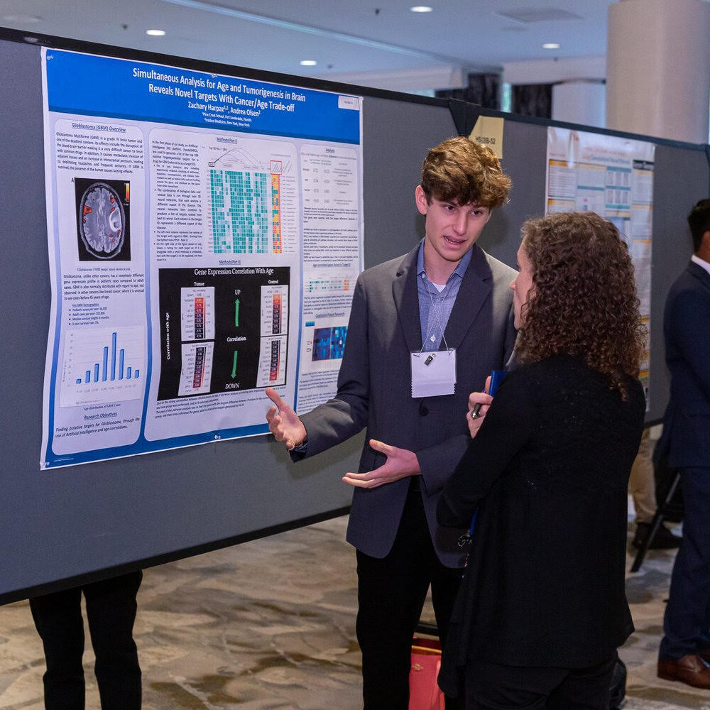
The good news here is that high school students, Zachary Harpaz, Andrea Olsen and Christopher Ren, and researchers Anastasia Shneyderman, Alexander Veviorskiy, Maria Dralkina, Simon Konnov, Olga Shcheglova, Frank W. Pun, Geoffrey Ho Duen Leung, Hoi Wing Leung, Ivan V. Ozerov, Alex Aliper, Mikhail Korzinkin, and Alex Zhavoronkov have recently made remarkable strides in the joint field of aging and glioblastoma research. The team used a generative artificial intelligence (AI) engine from Insilico Medicine (founded by Dr. Alex Zhavoronkov) called PandaOmics, to identify new therapeutic targets for both GBM and aging. On April 26, 2023, their research paper was published in Aging’s Volume 15, Issue 8, entitled, “Identification of dual-purpose therapeutic targets implicated in aging and glioblastoma multiforme using PandaOmics – an AI-enabled biological target discovery platform.”

Their Study

Andrea Olsen, a student at Sevenoaks School in Kent, UK, and CEO/co-founder of The Youth Longevity Association
Photo of Andrea Olsen, courtesy of Insilico Medicine

“[Glioblastoma multiforme] is one of the most horrible cancers because it has such a short survival time,” Andrea Olsen said. “Of course, it affects the brain and so affects the body because the brain is the control center of the entire body.”

Andrea Olsen, a student at Sevenoaks School in Kent, UK, and CEO/co-founder of The Youth Longevity Association, discovered her interest in neurobiology and technology while growing up in Oslo, Norway. In 2021, she started an internship at Insilico Medicine. Through her work with the researchers at Insilico Medicine, Olsen learned how to use AI to uncover new genetic targets that could be used to treat aging and cancer. Zachary Harpaz, a student at Pine Crest School in Fort Lauderdale, Florida, discovered his passion for biology after being introduced to the subject in 2020. He combined his passion for biology with his intrigue for computer science and AI to enter the field of aging research.

Zachary Harpaz, a student at Pine Crest School in Fort Lauderdale, Florida
Photo of Zachary Harpaz, courtesy of Insilico Medicine

“We wanted to find new putative targets for glioblastoma as well as aging—attacking them both at the same time,” explained Harpaz.

The researchers used a comprehensive approach to identify their targets. They split their data into three categories—young, middle-aged and senior—and mapped the importance of gene expression to survival. They analyzed 12 datasets and selected the genes that were overlapped in 11 of the 12 datasets. They also cross-referenced those genes with a recent study conducted by the researchers at Insilico Medicine in 2022 on putative targets for aging and certain diseases. 

PandaOmics

One of the most exciting aspects of their research was the use of PandaOmics. Typically, finding new drugs requires experts to comb through a myriad of data and conduct extensive research. With PandaOmics, AI quickly processes and analyzes the data to identify new therapeutic targets, reducing the time and resources required for drug development. These high school researchers used PandaOmics to screen datasets from the Gene Expression Omnibus repository (maintained by the National Center for Biotechnology Information) and discovered three new potential therapeutic targets for treating both aging and GBM.

The first target for glioblastoma and aging was CNGA3, which they selected after analyzing each gene through PandaOmics. They also analyzed negatively correlated genes and selected GLUD1 as the number one gene produced by PandaOmics. Finally, they cross-referenced genes highly correlated to aging with the previous 2022 study and selected SIRT1 as another potential therapeutic target. The students were excited by the findings. Before she interned with Insilico, Olsen said she didn’t realize that AI could be so helpful in finding completely new therapeutic targets. 

“For me, [working with Insilico] was an incredible opportunity to dive into the field of research, aging, longevity, and neuroscience. It really kick-started my entire career,” Olsen said.

Looking Ahead

Overall, the study conducted by the researchers and these high school students showcases the power of AI in drug discovery and highlights the potential for young researchers to make meaningful contributions to the field. Their findings could lead to the development of new therapies for glioblastoma and aging-related diseases. Other therapeutics identified through Insilico’s Pharma.AI platform—specifically for idiopathic pulmonary fibrosis and COVID-19—have already advanced to human clinical trials.

“I’m excited to continue my research into college, but I’m super grateful for this opportunity at Insilico. It allowed me to get a head start on learning how to conduct research, analyze data and use the coolest and most cutting edge AI in the drug development area,” Harpaz said. “That experience gave me an amazing head start and I’m super excited to continue that into college, and even after college.”

Click here to read the full research paper published by Aging.

AGING (AGING-US) VIDEOS: YouTube | LabTube | Aging-US.com

Aging is an open-access, peer-reviewed journal that has been publishing high-impact papers in all fields of aging research since 2009. These papers are available to readers (at no cost and free of subscription barriers) in bi-monthly issues at Aging-US.com.

Click here to subscribe to Aging publication updates.

For media inquiries, please contact media@impactjournals.com.

How Habitual Tea Drinking Impacts Brain Structure

In 2019, researchers conducted the first study to explore the effects of habitual tea drinking on system-level brain networks.

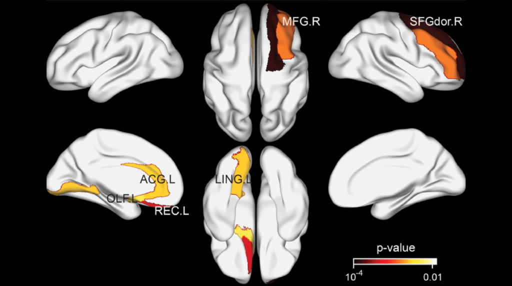
Figure 3. Brain regions exhibiting significant differences in structural nodal efficiency between the tea drinking group and the non-tea drinking group at the significance level of 0.01 (uncorrected) statistical evaluated by a permutation test.
Listen to an audio version of this article

After water, tea is the most popular beverage in the world. While many people enjoy tea for the flavor, aroma and caffeine boost, research suggests that there may be another reason to regularly drink this beverage: its effects on the brain. In 2019, researchers from Wuyi UniversityUniversity of EssexUniversity of Cambridge, and the National University of Singapore conducted the first study exploring the effects of tea on system-level brain networks. Their paper was published in Aging (Aging-US) Volume 11, Issue 11, and entitled, “Habitual tea drinking modulates brain efficiency: evidence from brain connectivity evaluation.”

“In this study, we comprehensively explored brain connectivity with both global and regional metrics derived from structural and functional imaging to unveil putative differential connectivity organizations between tea drinking group and non-tea drinking group.”

The Study

The subjects enrolled in this study were older adults (mean ≈ 70 years old) from residential communities in Singapore, without conditions or terminal illnesses (see Materials and Methods). Researchers initially recruited 93 participants, however, only 36 total participants (male = 6; female = 30) remained after adjusting for the strict study inclusion criteria. Researchers classified the remaining participants as “non-tea drinkers” or “tea drinkers” using complex composite test scores. The composite score included self-reports of multiple decades of weekly green tea, oolong tea, black tea, and coffee intake (see Materials and Methods). After screening, 15 participants were assigned to the tea-drinking group and 21 were assigned to the non-tea drinking group. (Coffee intake did not differ significantly between the two groups.)

Next, structural brain connectivity was compared between tea drinkers and non-tea drinkers. All 36 participants underwent MRI brain scans and both functional and structural networks were investigated from global and regional perspectives. The researchers found that participants in the tea-drinking group had more efficient structural organization. However, tea did not seem to have a significantly beneficial effect on global functional organization. As a result of tea drinking, hemispheric asymmetry in the structural connectivity network was observed, although it was not observed in the functional connectivity network. 

“In addition, functional connectivity strength within the default mode network (DMN) was greater for the tea-drinking group, and coexistence of increasing and decreasing connective strengths was observed in the structural connectivity of the DMN.”

Conclusion

The researchers found that tea drinkers had more efficient brain structure organization than non-tea drinkers. Studies have previously demonstrated that tea drinkers are less likely to develop dementia, and tea consumption has also been linked with better cognitive performance. The researchers note that these effects are due to tea’s contents of caffeine, L-theanine and polyphenols (catechins). Polyphenols are compounds found in plants, including tea leaves, and may help protect against oxidative damage. Previous studies have shown that tea polyphenols can cross the blood-brain barrier and may help improve brain function. 

While this study’s findings suggest that habitual tea drinking leads to better brain connectivity and efficiency in old age, the researchers were forthcoming about the limitations of their study. The sample size was limited and other substances, behaviors, habits, and environmental factors may have impacted the outcome of the study. 

“Our study offers the first evidence of the positive contribution of tea drinking to brain structure and suggests a protective effect on age-related decline in brain organisation.”

Click here to read the full research paper published by Aging (Aging-US).

AGING (AGING-US) VIDEOS: YouTube | LabTube | Aging-US.com

Aging (Aging-US) is an open-access journal that publishes research papers monthly in all fields of aging research and other topics. These papers are available to read at no cost to readers on Aging-us.com. Open-access journals offer information that has the potential to benefit our societies from the inside out and may be shared with friends, neighbors, colleagues, and other researchers, far and wide.

For media inquiries, please contact media@impactjournals.com.

Trending with Impact: Method Yields Cell-Type-Specific Brain Data

Researchers used a bioinformatics approach (ESHRD) that leverages gene expression data from brain tissue to derive cell-type specific alterations in Alzheimer’s disease.

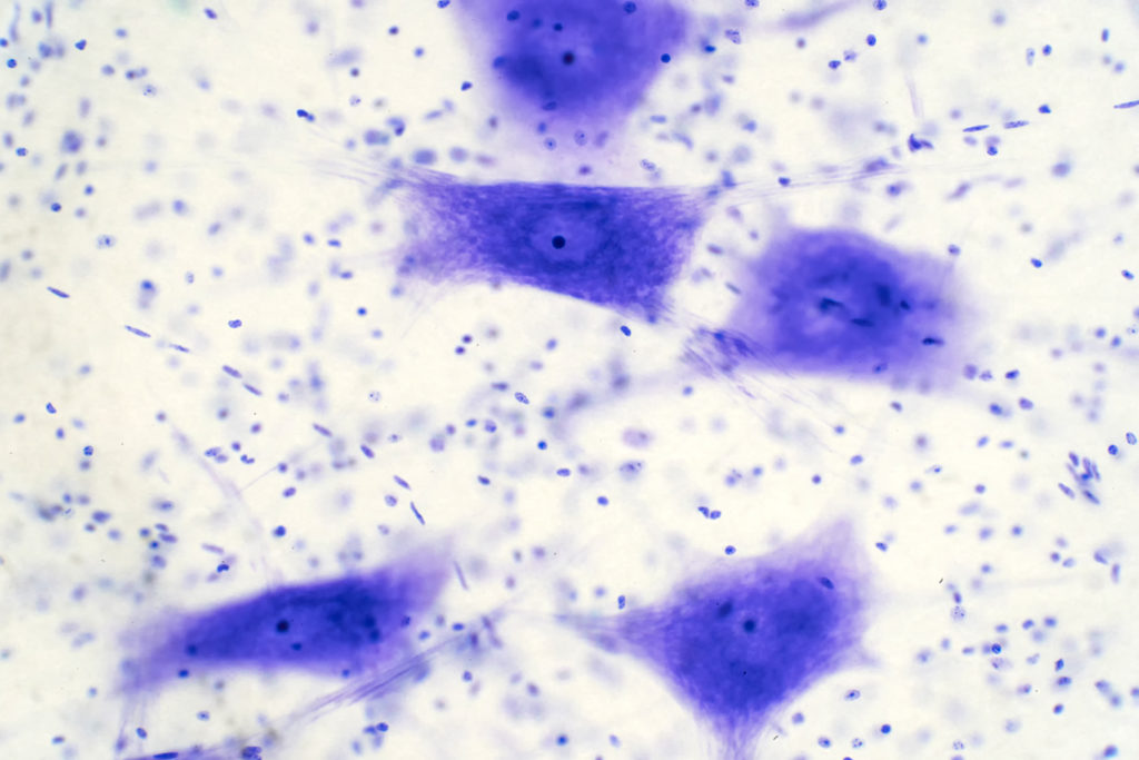
Neurons cells from the brain under the microscope.

The Trending with Impact series highlights Aging publications attracting higher visibility among readers around the world online, in the news, and on social media—beyond normal readership levels. Look for future science news about the latest trending publications here, and at Aging-US.com.

Listen to an audio version of this article

Cell-to-cell variability in the human brain is significantly heterogeneous. An abundance of differential brain cell types makes it laborious and expensive for researchers to generate single-cell gene expression data. While some studies use laser capture microdissection (LCM) and single-cell RNA sequencing (scRNA-Seq) to directly address the cellular heterogeneity in brain tissue, due to labor and cost, these studies generally have a small sample size and face power concerns. Most gene expression profiling studies of patients with Alzheimer’s disease (AD) are conducted post-mortem using brain tissue homogenates.

“Ultimately, the overall goal of gene expression profiling in AD is to understand the transcriptome changes in all major cell types of the brain in a well-powered approach that would facilitate the exploration of all the variables mentioned above.”

The need existed for a cost-effective bioinformatics approach to leverage expression profiling data from brain homogenate tissue to derive cell type-specific differential expression and pathway analysis results. In 2020, researchers from Columbia University Medical Center, The University of Sydney School of Medicine, University of Miami, and the Banner Sun Health Research Institute described an Enrichment Score Homogenate RNA Deconvolution (ESHRD) method for identifying alterations in the brain. They published a research paper in Aging’s Volume 12, Issue 5, entitled: “ESHRD: deconvolution of brain homogenate RNA expression data to identify cell-type-specific alterations in Alzheimer’s disease.” 

The Study

“We applied our approach to different gene expression datasets derived from brain homogenate profiling from AD patients and Non-Demented controls (ND) from 7 different brain regions.”

Researchers conducted brain region cell-specific pathway analysis and Gene Set Enrichment Analysis (GSEA). The team mapped and measured five different cell types in seven different brain regions. The cell types included: microglia, neuron, endothelial, astrocyte, and oligodendrocyte. Endothelial and oligodendrocyte are two cell types that are not easily examined in the brain and only very little gene expression data previously existed for Alzheimer’s disease.

“We conducted RNA expression profiling from both brain homogenates and oligodendrocytes obtained by LCM from the same donor brains and then calculated differential expression.”

The researchers used a dataset of Multiple System Atrophy (MSA) patients (n = 4) and controls (n = 5) to validate their ESHRD method. Homogenate, LCM, and scRNA-Seq results were compared using the ESHRD method. They also compared their findings to other research studies.

Results

“The ESHRD approach replicates previously published findings in neurons from AD patient brain specimens, and we extended our work to characterize novel AD-related changes in relatively unexplored cell types in AD, oligodendrocytes and endothelial cells.”

Neuronal, endothelial cells, and microglia were found to be the most represented “cell-specific” gene classes in patient brains with Alzheimer’s disease. Neuronal-specific genes were downregulated and enriched for synaptic processes. Endothelial genes were found to be upregulated in AD and enriched for angiogenesis and vascular functional processes.

“Differentially Expressed Genes (DEGs) we labeled as “mixed” represent the most prevalent class (73.4%), followed by DEGs labeled as microglia (6.6%), neuron (5.9%) and endothelial (5.7%). Astrocyte and oligodendrocyte labeled DEGs have a frequency of 3.6% and 3.1%, respectively.” 

Microglia showed different patterns of expression across the brain in multiple regions. They found that astrocyte genes were enriched in SLC transport and immune processes and oligodendrocytes were enriched for the Glycoprotein metabolism in Alzheimer’s disease.

Conclusion

“We demonstrate the ability of this approach to highlight known neuronal-specific changes in the AD brain and utilize it to identify novel changes in endothelial cells and oligodendrocytes, two cell types not easily examined in the brain and for which only minimal gene expression knowledge exists in AD.”

Click here to read the full study, published by Aging.

Aging is an open-access journal that publishes research papers monthly in all fields of aging research and other topics. These papers are available to read at no cost to readers on Aging-us.com. Open-access journals offer information that has the potential to benefit our societies from the inside out and may be shared with friends, neighbors, colleagues, and other researchers, far and wide.

Aging is a proud participant in the AACR Annual Meeting 2021 #AACR21

For media inquiries, please contact media@impactjournals.com.

  • Follow Us