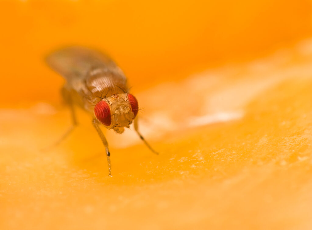
A new study by researchers from Osaka University’s Graduate School of Dentistry investigated cellular senescence in periodontal tissue and disease—identifying promising therapeutic targets for preventing periodontitis in the elderly.

The Trending With Impact series highlights Aging publications (listed by MEDLINE/PubMed as “Aging (Albany NY)” and “Aging-US” by Web of Science) that attract higher visibility among readers around the world online, in the news and on social media—beyond normal readership levels. Look for future science news about the latest trending publications here, and at Aging-US.com.
—
Repercussions of poor dental health aren’t limited to mere social stigmas. Poor dental health can impart serious consequences on an individual’s overall health. Periodontal disease broadly refers to any disease that affects the gums and the surrounding tissues that support the teeth, including the periodontal ligament (PDL) and alveolar bone. Periodontal disease can increase the risk of heart disease, stroke and diabetes by allowing bacteria to enter the bloodstream, causing inflammation and organ damage.
Periodontitis is a more advanced stage of periodontal disease. It is thought to be the most common infectious disease in the United States—affecting more than 40% of adults over 30 years old. Previous research has suggested that aging is a significant risk factor for periodontitis, although the underlying mechanisms are unclear.
“The direct cause of periodontitis is periodontopathic bacteria, while various environmental factors affect the severity of periodontitis. Previous epidemiological studies have shown positive correlations between aging and periodontitis. However, whether and how aging is linked to periodontal health and disease in biological processes is poorly understood.”
In a recent study, researchers Kuniko Ikegami, Motozo Yamashita, Mio Suzuki, Tomomi Nakamura, Koki Hashimoto, Jirouta Kitagaki, Manabu Yanagita, Masahiro Kitamura, and Shinya Murakami from Osaka University’s Graduate School of Dentistry aimed to elucidate the underlying mechanisms that contribute to aging-associated inflammation in periodontitis. On March 1, 2023, their new research paper was published in Aging (Aging-US) Volume 15, Issue 5, entitled, “Cellular senescence with SASP in periodontal ligament cells triggers inflammation in aging periodontal tissue.”
The Study
“In this study, we aimed to clarify the pathophysiological roles of cellular senescence in periodontal tissue and diseases.”
Previous studies have found that senescent cells can secrete senescence-associated secretory proteins (SASP) that induce inflammation and impair wound healing in some chronic diseases. The existence of senescent cells in periodontal tissue and diseases, however, has yet to be clarified. In this study, the researchers investigated cellular senescence and SASP in aging periodontal tissue. The team aimed to uncover the mechanism by which cellular senescence and SASP trigger inflammation in periodontal tissue and to identify potential therapeutic targets for this disease.
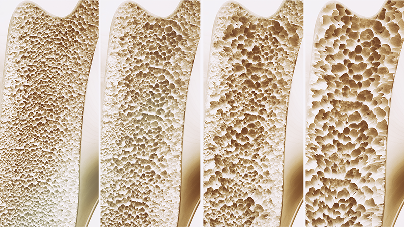
To investigate the role of cellular senescence in periodontitis, the researchers analyzed periodontal tissue in young and aged mice. Alveolar bone volume was compared in the young and aged mice, and beta-galactosidase (β-gal) staining was performed. They found bone resorption in aged mice and many senescence-associated (SA) β-gal-positive cells in their periodontal tissue, leading to inflammation and breakdown of alveolar bone. Very few SA β-gal-positive cells were found in young mouse tissues.
Next, the researchers worked with cells in vitro, primary human periodontal ligament (HPDL) cells, and induced cellular senescence through serial passaging (replicative senescence). The growth rate of HPDL cells gradually reduced, and they reached irreversible cell growth arrest, indicating the induction of cellular senescence. Morphological changes were observed through phalloidin staining. The team found that around 70% of aged HPDL cells were positive for SA β-gal, while less than 10% of young HPDL cells were positive. Morphological changes showed that aged HPDL cells had an enlarged and “spread” cell shape compared to young HPDL cells. Flow cytometry analysis confirmed an increase in cell size and granularity of aged HPDL cells compared to young HPDL cells.
TEM analysis showed that senescent cells exhibit metabolic changes and irregularly shaped mitochondria with disrupted cristae and increased accumulation of ROS, which suggest damage and failure of the redox balance. Importantly, the researchers found that the intrinsic inflammation state of aged PDLs was higher than in young PDLs, and susceptibility to bactericidal pathogens (but not inflammatory cytokines) was low in aged PDLs. Additionally, the team observed an age-dependent upregulation of microRNA (miR)-34a in HPDL cells.
“Thus, miR-34a and senescent PDL cells might be promising therapeutic targets for periodontitis in elderly people.”
Summary & Conclusion
In conclusion, poor dental health can have serious implications on an individual’s overall health, as periodontal disease can increase the risk of heart disease, stroke and diabetes. Aging is a significant risk factor for periodontitis, which is thought to be the most common infectious disease in the United States. A recent study by researchers from Osaka University’s Graduate School of Dentistry aimed to elucidate the underlying mechanisms that contribute to aging-associated inflammation in periodontitis. The study found that senescent cells in periodontal tissue secrete SASP that induce inflammation. The researchers identified potential therapeutic targets for periodontitis and suggest that elimination of senescent PDL cells or suppression of the miR-34a-dependent SIRT1-NF-κB axis may be an attractive therapeutic strategy to prevent periodontitis in humans as we age.
“To the best of our knowledge, this is the first study to identify: 1) the potential for senescent PDL cells to induce inflammation of periodontal tissue, and 2) a miRNA-dependent molecular mechanism of SASP in senescent PDL cells.”
Click here to read the full research paper published by Aging.

AGING (AGING-US) VIDEOS: YouTube | LabTube | Aging-US.com
—
Aging is an open-access, peer-reviewed journal that has been publishing high-impact papers in all fields of aging research since 2009. These papers are available to readers (at no cost and free of subscription barriers) in bi-monthly issues at Aging-US.com.
Click here to subscribe to Aging publication updates.