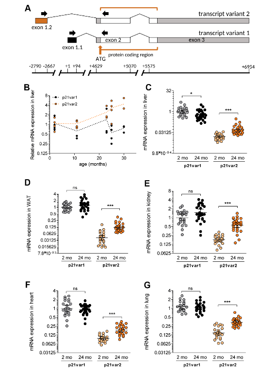
In a priority research paper published by Aging-US in January of 2022, researchers investigated aged muscle stem cells and their ability to sense and respond to mechanical cues.
The Trending With Impact series highlights Aging (Aging-US) publications that attract higher visibility among readers around the world online, in the news, and on social media—beyond normal readership levels. Look for future science news about the latest trending publications here, and at Aging-US.com.
—
IIs muscle wasting a fate humans can avoid, or will the problem of aging-related muscle loss only be resolved when the mystery of aging is solved? Researchers—from Vrije Universiteit Amsterdam, University of Amsterdam, Sorbonne Université, Amsterdam University Medical Center VUmc, Université Catholique de Louvain, KU Leuven, and Institut NeuroMyoGène—conducted a study aimed at elucidating whether muscle stem cells are inherently impaired by the aging process in their ability to sense and respond to mechanical cues. Their priority research paper was published in January of 2022 on the cover of Aging (Aging-US) Volume 14, Issue 1, and entitled, “Reduced growth rate of aged muscle stem cells is associated with impaired mechanosensitivity.”
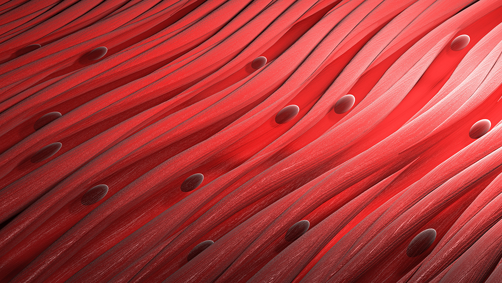
Muscle Stem Cells
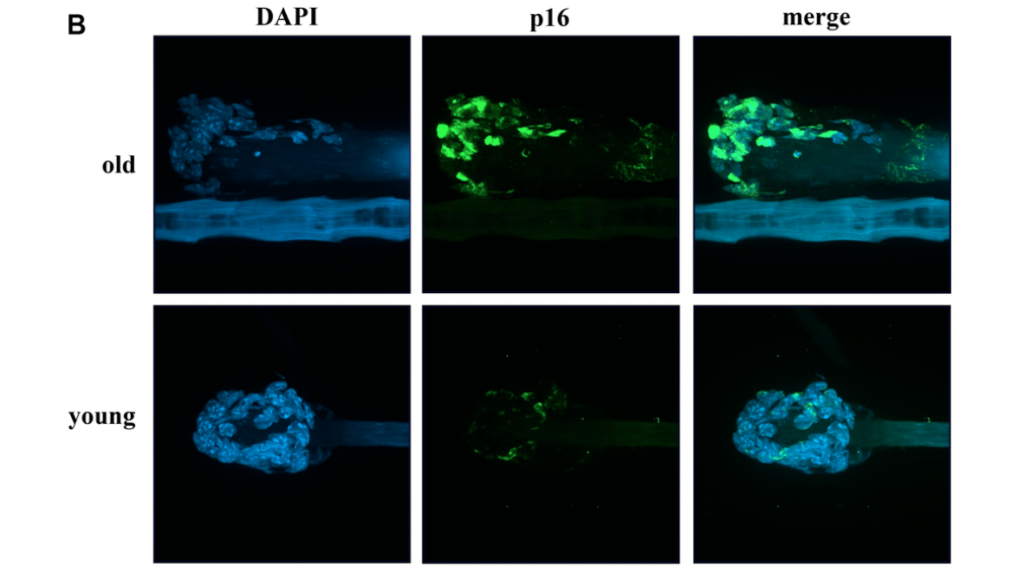
Muscle stem cells (MuSCs) are stem cells located within skeletal muscle tissues. MuSCs function to repair Muscle stem cells (MuSCs) are stem cells located within skeletal muscle tissues. MuSCs function to repair damaged myofibers and give rise to new skeletal muscle cells. These self-renewing stem cells are involved in muscle growth, repair and regeneration. As we age, MuSCs decline in number and lose their potential to regenerate damaged myofibers, leading to sarcopenia. The researchers in this study hypothesized that the responsiveness of aged MuSCs is impared by the aging process both physically and mechanically.
“We postulated that aged MuSCs are intrinsically impaired in their responsiveness to omnipresent mechanical cues through alterations in MuSC morphology, mechanical properties, and number of integrins, culminating in impaired proliferative capacity.”
The Study
The researchers assessed whether aged MuSCs become impaired in their ability to proliferate, respond to pulsating fluid shear stress (PFSS) mechanical loading, maintain focal adhesion number and/or size after mechanical loading, and in their ability to express the protein-coding gene Integrin Subunit Alpha 7 (ITGA7).
“Integrins are transmembrane protein receptors that connect MuSCs to the ECM [extracellular matrix] components and are part of focal adhesions [51].”
Young MuSCs (2 months) and aged MuSCs (22 months) were isolated from male mice. Fluorescence-activated cell purification was carried out and cells were cultured. To measure proliferation, images were captured of the cell cultures every 24 hours. Images were also taken pre- and post-PFSS to determine the number of young and aged MuSCs detached from the culture media (focal adhesion) as a result of PFSS treatment. Since nitric oxide (NO) is known to play a role in MuSC activation and muscle regeneration, NO analysis was conducted to measure NO production. To determine MuSC morphology, the researchers carried out immunohistochemistry staining. They also measured MuSC stiffness, deformation, gene expression, and RNA isolation and reverse transcription.
Compared to young MuSCs, the researchers found aged MuSCs had impaired growth. Their results showed that IL-6 gene expression was lower in aged MuSCs, which suggested that aged MuSCs were intrinsically altered in the signaling pathways governing proliferation and MuSC function. Aged MuSCs showed an increase in cell volume and reduced cell adhesion after mechanical loading. NO levels in young and aged MuSCs were similar, and PFSS in both cultures resulted in similar increases in NO production. The researchers found decreased ITGA7 expression and reduced pPXN clusters (focal adhesion formation) were involved in altered MuSC function with age. High YAP nuclear localization was found in aged MuSCs, as well as reduced mechanosensitivity.
“Aged MuSCs were less sensitive to shear forces and showed upregulation of less genes, suggesting that the decreased mechanosensitivity was due to decreased integrin protein expression, i.e. ITGA7, ITGA5, and ITGB5, and focal adhesion number.”
Conclusion
The results from this study found that aged MuSCs were intrinsically impaired in their growth rate due to decreased ITGA7 expression and diminished focal adhesion formation. These changes coincided with increased cell volume, decreased MuSC adhesion, altered mechanosensitivity, changed YAP signaling and decreased expression of several genes (including cell cycle genes). The researchers suggest that ITGA7 and pPXN may be potential therapeutic targets to improve aged MuSC function.
“As an implication, a possible therapeutic option could be restoration ITGA7 and focal adhesion number in aged MuSCs, which may help to restore MuSCs adhesion to their niche as well as growth rate of these cells.”
Click here to read the full priority research paper published by Aging (Aging-US).

AGING (AGING-US) VIDEOS: YouTube | LabTube | Aging-US.com
—
Aging (Aging-US) is an open-access journal that publishes research papers bi-monthly in all fields of aging research. These papers are available to read at no cost to readers on Aging-us.com. Open-access journals offer information that has the potential to benefit our societies from the inside out and may be shared with friends, neighbors, colleagues, and other researchers, far and wide.
For media inquiries, please contact media@impactjournals.com.