Behind the Study: Dr. Andrei Gudkov

Aging Editorial Board member Andrei V. Gudkov, PhD, DSci, discusses his 2017 research paper published by Aging, entitled, “p16(Ink4a) and senescence-associated β-galactosidase can be induced in macrophages as part of a reversible response to physiological stimuli.”

Researchers explain their studies that were published in Aging

Behind the Study is a series of transcribed videos from researchers elaborating on their recent oncology-focused studies published in Aging. A new Behind the Study is released each Monday. Visit the Aging YouTube channel for more insights from outstanding authors.

Greetings. My name is Andrei Gudkov. I am working in Roswell Park Cancer Institute, designated cancer center located in Buffalo, New York. I am Senior Vice President for Basic Science and chair of Department of Cell Stress Biology. My research is focused on understanding of the mechanisms of deregulation of a variety of stress response pathways in cancer cells as well as in normal cells in relation to cancer origin, progression, or engraftment and trying to use the information which we are generating during this research to come up with new types of treatment of cancer or cancer prevention.

Recently, our interests have significantly switched towards studying of the mechanisms of aging in its relations to cancer, since, as we all know, both conditions are closely connected. During the last, probably 20 years, one of the central theories of aging in mammals has been evolving towards connection between chronic sterile inflammation, which is accumulating in tissues with age of a mammal, including humans, with systemic decline in regeneration capabilities, in function of organs and tissues, and increasing risk of major diseases, altogether known as aging-related diseases. And the source of this inflammation, its origin, has been the central focus of studies of many.

During last couple of years, the dominating opinion in the field is about the central role of senescent cells, cells which chose to stay irreversibly growth-arrested in response to DNA damage, which they acquire during their life. And, through that, change their phenotype in more significant way than just growth arrest, acquiring the ability to secrete a spectrum of pro-inflammatory factors.

These senescent cells, which initially were defined as such in tissue culture experiments, eventually were proclaimed to be the main suspects in their putative role of inflammation creators in aging organism. This idea has become really popular, especially following a series of brilliant works coming from number of laboratories, in which senescent cells were detected in vivo in mice and mouse models. And when these mice were treated with agents which eradicated these senescent cells, numerous signs of rejuvenation were observed.

I’m talking about the first paper of that kind appeared in 2011, Mayo Clinic, and the group led by Jim Kirkland and Jan van Deursen and a series of follow-up papers with similar results. In general, the idea of putting senescent cells in the position of the key sources of sterile chronic inflammation associated with aging came from Judy Campisi, who has provided the most important discoveries in that field.

Well, this theory is extremely appealing for many reasons. First, it is very well supported by evidence. Indeed, senescent cells, when they turn into senescents in culture, switch their phenotype into, so-called, SASPs, and that’s an associated secretory phenotype, the state in which cells continuously secrete pro-inflammatory factors. Second, these cells appear in culture as a result of serial passaging resembling aging. And, therefore, this link became kind of natural between aging and senescent cells. The presumption was that certain cells in the body who used up the number of divisions they can go through before they reach this state may be increasing with age and, therefore, these cells accumulate.

Each of them may become the source of sterile inflammation. Each single one provides a very weak signal, but, when they accumulate altogether, the impact may become significant and translated into pathological conditions. So recently, there were very few – and, even now, it is like that – very few biomarkers of senescent cells, none of which is very reliable because every single biomarker is kind of promiscuous and is not universally selective for senescent cells.

Among these biomarkers, two have been most popular. One is high level of expression of, so-called, senescent-associated senescent-associated beta-galactosidase, which can be detected chemically in fixed cells and tissues which undergo staining, including X-gal, which turn beta-galactosidase reaction into the blue dye under conditions which is not optimal for endogenous beta-galactosidases mammalian cells at low pH. And, under these conditions, the background beta-gal activity of normal cells is practically not seen and senescent cells become brightly visible. So this reaction, which unfortunately requires a cell… It can not be done on paraffin-embedded sections and require preservation of the enzymatic activity and, therefore, is available, mostly, on the frozen sections or in cells in culture… has been used very, very frequently. And in many papers, it has been just the only assay which was used for detection of so-called senescent cells.

Figure 1. Induction of p16Ink4a and SAβG in macrophages does not require p53.

The other biomarker, which resulted from a detailed analysis of promoters which are active selectively in senescent cells is the gene encoding cyclin-dependent kinase inhibitor p16. And the genes name is INK4a. In fact, this promoter of this gene is frequently upregulated in senescent cells, and it has relatively low background in other cells of the organism.

Again, p16 activation is not limited to senescent cells and, moreover, not every senescent cells has elevated p16, but that’s the best we have as of today. That is why, whenever the investigators want to create a mouse model in which they could have the desirable gene expressed exclusively in senescent cells, they use p16 promoter. And there are several mouse models; I’m aware of three in which reported constructs were put under p16 promoter. And the claim was that, when these reporters become obviously expressed in mouse tissues, that was interpreted as accumulation of senescent cells. Also, one can put under this promoter the gene which enables selective eradication of cells with this expression, and, therefore, there is an opportunity to selectively kill such cells. Again, this can be interpreted as a selective eradication of senescent cells.

Using these models, two groups of investigators claim that eradication of senescent cells in aged mice led to substantial demonstration of signs of rejuvenation and, in one case, with increased lifespan. Well, obviously, these data not only provided a very powerful support for the theory about the role of senescent cells in aging but also provided the proof of concept for development of pharmacological approaches to anti-aging treatment and treatment of conditions which lead to the high risk of development of age-related diseases, including cancer.

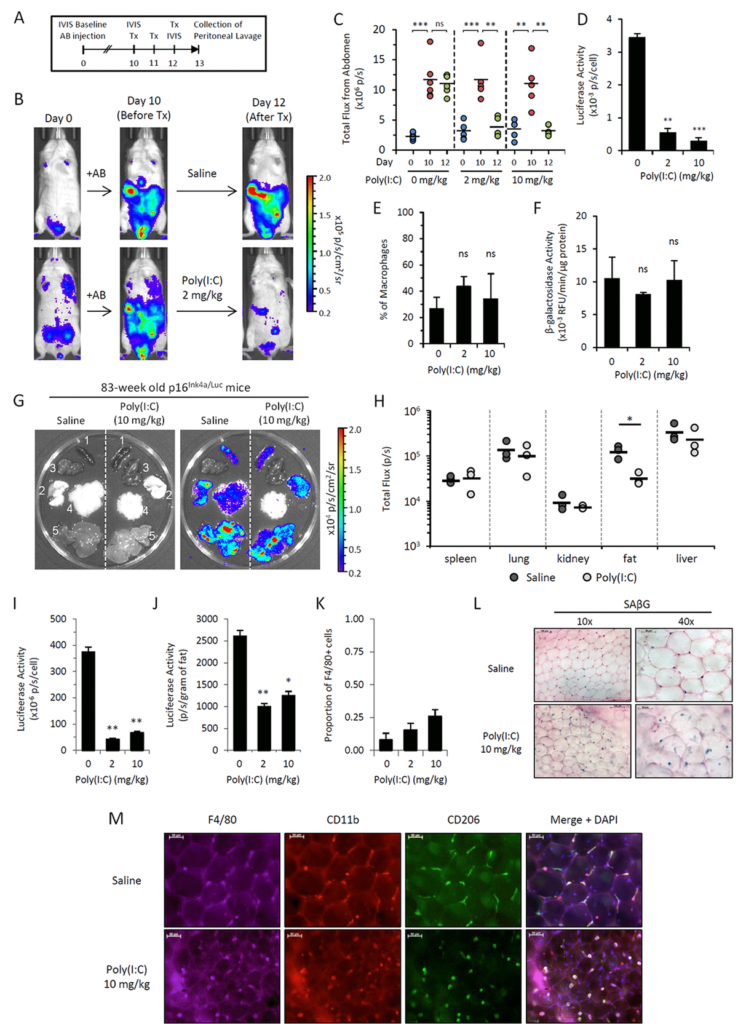
We obtained such mice in our laboratory, and we have been working with them during last couple of years. The mice we are using are coming from the laboratory of Norman Sharpless from North Carolina. And they have a luciferase reporter gene, which is substituting one of the alleles of p16 and, thereby, being expressed from the p16 promoter. We were pleased to see that these mice accumulate p16-driven luciferase-positive cells detected by in vivo imaging during their lives, which, actually, very well fit the senescent cell theory in their accumulation during life.

However, we were very surprised not seeing accumulation of these cells following total-body radiation or treatment with other genotoxic conditions, which, supposedly, should create lots of senescent cells. We also were puzzled that we were unable to see activation of p16-driven luciferase when we take tissues from these mice and isolated mesenchymal cells from these tissues in vitro and then turn them into senescents, and we failed to see activation of luciferase.

Again, all this together stimulated us to look at the nature of p16-positive cells in these mice and determined their nature, their origin, and their fate in vivo. We started from following the consequences of injection of cells, which would turn into senescents in vitro following injection in vivo into mice. And we labeled cells. We made cell senescents in culture by gum radiation. Then, we injected them intraperitoneal, subcutaneously, into mice. And we looked for their presence by monitoring the label which they were marked with.

Well, it appears that these labeled cells – their traces are disappearing quite quickly, and, within a few days, there are none left in the mice. However, if you put normal cells of similar origin, they actually last much longer. That was the first indication that there may be a mechanism of selective eradication of senescent cells in the body. To check this mechanism and one of our hypotheses was that this mechanism is associated with physical attack of some cells of immunity against senescent cells, and there’s supposed to be innate immunity because it’s happening immediately without any education over the organism.

Figure 5. Poly(I:C) abrogates elevated p16Ink4a expression in two independent in vivo models. 

We use a trick in which we embedded senescent cells created in vitro into algenate beads, small spheres consisting of a polymer, which enables to keep cells alive inside them, does not interfere with acquisition of nutrients and oxygen by the cells, but prevents any attack against the cells from any immunocytes. When we took these beads filled with senescent cells and put them in peritoneal cavity of mice, we were pleased to see that they are lasting four weeks without significant death, indicating that senescent cells, who disappear if they are injected without protective beads, are indeed killed by some, so far, unknown mechanism.

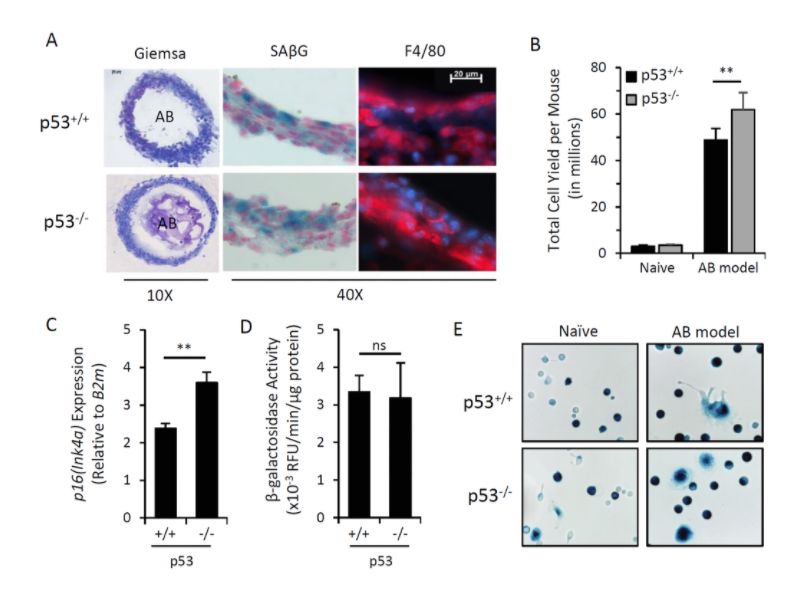
In order to identify the executors of senescent cells, we put these beads filled with senescent cells as bait inside, very peritoneal cavity of normal mice, and two weeks later, we pulled them out and analyzed who was accumulating in terms of how cells around these beads in lavage liquid, as well as in the capsule, which was formed around every bead.

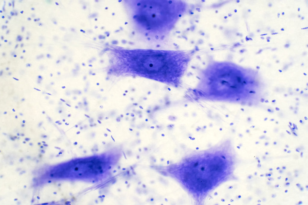
Our results brought us to a very important and quite striking observational. The major part of the cells, which was so in these beads as well as in the lavage, appeared to be cells with macrophageal markers on them, which appeared to be bright fluorescence, meaning that they have activated p16, and also positive for beta-gal staining conducted under conditions we are using to reveal senescent cells. So we had to conclude that senescent cells put in the beads attract, probably, by the products of their secretion special subtypes of immune cells, significant proportion of which become reprogrammed to start expressing two biomarkers which people have been using to distinguish senescent cells.

We studied these macrophages in detail, and, after we published our first paper in which we describe this phenomenon, we published a second one, also in Aging, where their properties were described in further details. And we confident that these are bonafide macrophages, not only because they have have biomarkers, they have surface antigen specific for macrophages, but also they are capable of phagocytosis and, moreover, they can be selectively killed by liposome-embedded clodronate, a poison which only kills cells capable of phagocytosis. This killing could be done both in vitro and in vivo when you inject liposomal clodronate inside mice.

So, as far as the presence of these cells in the body of those mice which are not embedded with algenate beads with senescent cells, today, we are confident that these macrophages are accumulating in subcutaneous fat of aged mice in large numbers. And, again, they express biomarkers of macrophages that can be selectively eradicated by clodronate.

So, altogether, it means that the cells which become p16-positive vivo, not necessarily our senescent cells – our operations does not disprove that the signal which we and other investigators are seeing in these mice and increasing with age is not associated with senescent cells. So, potentially, certain proportion of cells we see are, indeed, senescent. However, we are confident that significant part of the signal goes from macrophages, which can be induced into the phenotype associated with expression of both senescent markers when they’re exposed to senescent cells. What is also interesting that this phenotype is reversible. And, in our second paper, we provide a number of physiological stimuli which can either stimulate or suppressed acquisition of this phenotype by macrophages.

All this, together, provides a very interesting step forward in evolution of the theory of aging associated with accumulation of certain specific cell types, contributing to the sterile inflammation occurring in tissues. Today, we can say that those cells which we claim to be the main source of that are not necessarily senescent, but also can be immunocytes who share with senescent cells some of their properties but are not senescent by nature and simply reprogrammed macrophages.

What is the relative impact of these macrophages versus senescent cells towards the process of aging is a very important question, not only from a theoretical standpoint, but also from practical standpoint because, from the time when senescent cells were claimed to be the key players of aging, there have been a substantial effort in the field in generating and testing senolytic compounds, drugs, emerging drugs, which potentially can have anti-aging effect due to eradication of senescent cells from the body.

Whether senolytic compounds would, indeed, solve the issue because, presumably, they will eliminate only a part of the p16-positive cells. To what extent, we need to redirect our attention to the senescent cell-associated macrophages as potential alternative source of secreted factors is an open question. And these are the questions which we are trying to address in our ongoing work, which stems from these observations. Thank you.

Click here to read the full study published in Aging.

Aging is an open-access journal that publishes research papers monthly in all fields of aging research and other topics. These papers are available to read at no cost to readers on Aging-us.com. Open-access journals offer information that has the potential to benefit our societies from the inside out and may be shared with friends, neighbors, colleagues, and other researchers, far and wide.

For media inquiries, please contact media@impactjournals.com.

Aging is a proud participant in the AACR Annual Meeting 2021 #AACR21

Trending with Impact: Method Yields Cell-Type-Specific Brain Data

Researchers used a bioinformatics approach (ESHRD) that leverages gene expression data from brain tissue to derive cell-type specific alterations in Alzheimer’s disease.

Neurons cells from the brain under the microscope.

The Trending with Impact series highlights Aging publications attracting higher visibility among readers around the world online, in the news, and on social media—beyond normal readership levels. Look for future science news about the latest trending publications here, and at Aging-US.com.

Listen to an audio version of this article

Cell-to-cell variability in the human brain is significantly heterogeneous. An abundance of differential brain cell types makes it laborious and expensive for researchers to generate single-cell gene expression data. While some studies use laser capture microdissection (LCM) and single-cell RNA sequencing (scRNA-Seq) to directly address the cellular heterogeneity in brain tissue, due to labor and cost, these studies generally have a small sample size and face power concerns. Most gene expression profiling studies of patients with Alzheimer’s disease (AD) are conducted post-mortem using brain tissue homogenates.

“Ultimately, the overall goal of gene expression profiling in AD is to understand the transcriptome changes in all major cell types of the brain in a well-powered approach that would facilitate the exploration of all the variables mentioned above.”

The need existed for a cost-effective bioinformatics approach to leverage expression profiling data from brain homogenate tissue to derive cell type-specific differential expression and pathway analysis results. In 2020, researchers from Columbia University Medical Center, The University of Sydney School of Medicine, University of Miami, and the Banner Sun Health Research Institute described an Enrichment Score Homogenate RNA Deconvolution (ESHRD) method for identifying alterations in the brain. They published a research paper in Aging’s Volume 12, Issue 5, entitled: “ESHRD: deconvolution of brain homogenate RNA expression data to identify cell-type-specific alterations in Alzheimer’s disease.” 

The Study

“We applied our approach to different gene expression datasets derived from brain homogenate profiling from AD patients and Non-Demented controls (ND) from 7 different brain regions.”

Researchers conducted brain region cell-specific pathway analysis and Gene Set Enrichment Analysis (GSEA). The team mapped and measured five different cell types in seven different brain regions. The cell types included: microglia, neuron, endothelial, astrocyte, and oligodendrocyte. Endothelial and oligodendrocyte are two cell types that are not easily examined in the brain and only very little gene expression data previously existed for Alzheimer’s disease.

“We conducted RNA expression profiling from both brain homogenates and oligodendrocytes obtained by LCM from the same donor brains and then calculated differential expression.”

The researchers used a dataset of Multiple System Atrophy (MSA) patients (n = 4) and controls (n = 5) to validate their ESHRD method. Homogenate, LCM, and scRNA-Seq results were compared using the ESHRD method. They also compared their findings to other research studies.

Results

“The ESHRD approach replicates previously published findings in neurons from AD patient brain specimens, and we extended our work to characterize novel AD-related changes in relatively unexplored cell types in AD, oligodendrocytes and endothelial cells.”

Neuronal, endothelial cells, and microglia were found to be the most represented “cell-specific” gene classes in patient brains with Alzheimer’s disease. Neuronal-specific genes were downregulated and enriched for synaptic processes. Endothelial genes were found to be upregulated in AD and enriched for angiogenesis and vascular functional processes.

“Differentially Expressed Genes (DEGs) we labeled as “mixed” represent the most prevalent class (73.4%), followed by DEGs labeled as microglia (6.6%), neuron (5.9%) and endothelial (5.7%). Astrocyte and oligodendrocyte labeled DEGs have a frequency of 3.6% and 3.1%, respectively.” 

Microglia showed different patterns of expression across the brain in multiple regions. They found that astrocyte genes were enriched in SLC transport and immune processes and oligodendrocytes were enriched for the Glycoprotein metabolism in Alzheimer’s disease.

Conclusion

“We demonstrate the ability of this approach to highlight known neuronal-specific changes in the AD brain and utilize it to identify novel changes in endothelial cells and oligodendrocytes, two cell types not easily examined in the brain and for which only minimal gene expression knowledge exists in AD.”

Click here to read the full study, published by Aging.

Aging is an open-access journal that publishes research papers monthly in all fields of aging research and other topics. These papers are available to read at no cost to readers on Aging-us.com. Open-access journals offer information that has the potential to benefit our societies from the inside out and may be shared with friends, neighbors, colleagues, and other researchers, far and wide.

Aging is a proud participant in the AACR Annual Meeting 2021 #AACR21

For media inquiries, please contact media@impactjournals.com.

Complex Drug Screenings Show Anti-Aging Potential in Natural Compounds

In an effort to mimic metformin and rapamycin, researchers used powerful screening methods to analyze over 800 natural compounds to assess their anti-aging potential and safety profile.

Modern Medical Research Laboratory. Scientific Lab, Drug Engineering Center Full of High-Tech Equipment, Computer Screen Showing DNA concept, Technology for Vaccine Development.

The Top-Performer series highlights papers published by Aging that have generated a high Altmetric attention score. Altmetric scores, located at the top-left of trending Aging papers, provide an at-a-glance indication of the volume and type of online attention the research has received.

Read Aging’s Top 100 Altmetric papers.

Listen to an audio version of this article

By 2030, one in five Americans will age over 65 years old in the United States. As a society, an even larger aging population is fast on our heels. With age comes wisdom, and unfortunately, so does a number of costly and devastating diseases, including cancer, cardiovascular disease, Alzheimer’s disease, and Type II diabetes.

Researchers are currently working to mitigate the upcoming burden for this expanding population by developing anti-aging and anti-cancer drugs, and other geroprotective interventions that could extend healthspan, lower disease rates, and maintain productivity. However, the slow and expensive process of gaining approval for new potential pharmaceutical and nutraceutical interventions is historically arduous and prone to failure—especially when it comes to anti-aging and longevity research.

“Even if successful, to be used preventatively, anti-aging drugs face extraordinarily high safety and efficacy standards for approval [9].”

In 2017, researchers from the United States’ Insilico Medicine, Inc. and Life Extension, the United Kingdom’s Biogerontology Research Foundation, Canada’s Queen’s University, and Russia’s Russian Academy of Sciences, worked together to test a strategy to accelerate the development of safe, wide-scale anti-aging nutraceuticals. Their study was published in Aging’s Volume 9, Issue 11, and entitled, “Towards natural mimetics of metformin and rapamycin.” To date, this top-performing research paper has generated an Altmetric Attention score of 127.

Metformin and Rapamycin

“One strategy to hasten the process has been the repurposing of existing, FDA-approved drugs that show off-label anti-cancer and anti-aging potential [10,11], and at the top of that list are metformin and rapamycin, two drugs that mimic caloric restriction [12].”

Metformin and rapamycin have already been FDA approved for use in renal transplants, Type II diabetes, and metabolic syndrome. These two drugs are both mTOR inhibitors which, through numerous research studies, have shown pleiotropic effects exhibiting multiple anti-aging, anticancer, and anti-cardiovascular disease benefits. However, some adverse side effects pertaining to extended use have made it so these two interventions (used alone) are unable to move forward for wide-scale preventative use.

“Taken together, rapamycin and metformin are promising candidates for life and healthspan extension; however, concerns of adverse side effects have hampered their widescale adoption for this purpose.”

Although there are some adverse side effects, the chemical structures of metformin and rapamycin should not be ignored. These two drugs can be analyzed, and even mimicked, to develop new, safer interventions to prevent and treat age-related diseases. The researchers in this study initiated an effort to identify nutraceuticals as safe, natural alternatives to metformin and rapamycin drugs. 

“Nutraceuticals have received considerable attention in recent years for potential roles in preventing or treating a number of age-related diseases [88].”

The Study

“Our work is done entirely in silico and entails the use of metformin and rapamycin transcriptional and signaling pathway activation signatures to screen for matches amongst natural compounds.” 

Test compounds were selected based on the natural compounds listed in the UNPD and Library of Integrated Network‐based Cellular Signatures (LINCS) datasets. Gene‐ and pathway‐level signatures of metformin and rapamycin were mapped and screened for matches against the over 800 natural compounds chosen. The team used conventional statistical methods, pathway scoring-based methods, and training of deep neural networks for signature recognition. Researchers applied several bioinformatic approaches and deep learning methods, including the Oncofinder, Geroscope, and in silico Pathway Activation Network Decomposition Analysis (iPANDA). The iPANDA extracts robust, biologically relevant pathway activation signatures from the data. 

“In an application of these methods, we focused on mimicry of metformin and rapamycin, seeking nutraceuticals that could preserve their anti-aging and disease-preventive potential while being better suited for wide-scale prophylactic use.”

Results and Conclusion

“The analysis revealed many novel candidate metformin and rapamycin mimetics, including allantoin and ginsenoside (metformin), epigallocatechin gallate and isoliquiritigenin (rapamycin), and withaferin A (both). Four relatively unexplored compounds also scored well with rapamycin.”  

Their initial list of over 800 natural compounds was condensed to a shortlist of candidate nutraceuticals that showed similarity to metformin and rapamycin and had low adverse effects. 

“This work revealed promising candidates for future experimental validation while demonstrating the applications of powerful screening methods for this and similar endeavors.”

Click here to read the full research paper, published by Aging.

Aging is an open-access journal that publishes research papers monthly in all fields of aging research and other topics. These papers are available to read at no cost to readers on Aging-us.com. Open-access journals offer information that has the potential to benefit our societies from the inside out and may be shared with friends, neighbors, colleagues, and other researchers, far and wide.

Aging is a proud participant in the AACR Annual Meeting 2021 #AACR21

For media inquiries, please contact media@impactjournals.com.

Trending with Impact: Epigenetic Age Decreased in Diet & Lifestyle Study

Researchers conducted an eight-week study on diet and lifestyle among a small cohort of 43 male participants between the ages of 50 and 72.

Happy senior couple buying fresh food at the market

The Trending with Impact series highlights Aging publications attracting higher visibility among readers around the world online, in the news, and on social media—beyond normal readership levels. Look for future science news about the latest trending publications here, and at Aging-US.com.

Listen to an audio version of this article

In addition to the well-known personal and social costs of aging, the economic costs of aging are also considerably high. Research finds that investing in delaying aging is much more cost-effective than disease-specific spending. A study found that if Americans as a whole delayed their aging by 2.2 years (while extending healthspan), economic savings over 50 years could amount to a cumulative $7 trillion.

“The growing health-related economic and social challenges of our rapidly aging population are well recognized and affect individuals, their families, health systems and economies.”

Across three countries (the United States, Canada, and Israel), researchers from the Institute for Functional Medicine, American Nutrition Association, National University of Natural Medicine, Ariel University, McGill University, and the University of California, conducted a new pilot study on the effects that diet and lifestyle intervention have on aging among healthy males between the ages of 50 and 72. This research paper was published in Aging’s Volume 13, Issue 7, and entitled, “Potential reversal of epigenetic age using a diet and lifestyle intervention: a pilot randomized clinical trial.”

The Study

The researchers organized a cohort of 43 healthy adult males between the ages of 50 and 72. Half of the participants (n=21) completed an eight-week treatment program, and the other half (control group=22) received no intervention. Interventions within the treatment program included regimented diet, sleep, exercise, relaxation guidance, and supplemental probiotics and phytonutrients. Prior to the treatment program, participants were enrolled in a preliminary education week to become acquainted with the researchers’ prescribed dietary and lifestyle interventions.

“To our knowledge, this is the first randomized controlled study to suggest that specific diet and lifestyle interventions may reverse Horvath DNAmAge (2013) epigenetic aging in healthy adult males.”

Diet Prescription

Researchers prescribed the participants with mostly (not entirely) plant-based diet instructions to consume measured portions of liver, eggs, dark leafy greens, cruciferous vegetables, colorful vegetables (excluding white potatoes and sweetcorn), beets, pumpkin seeds (or pumpkin seed butter), sunflower seeds (or sunflower seed butter), methylation adaptogens, berries, rosemary, turmeric, garlic, green tea, oolong tea, animal protein, and low glycemic fruit. They were prescribed two daily doses of PhytoGanix®, which is a combination of organic vegetables, fruits, seeds, herbs, plant enzymes, prebiotics, and probiotics. A daily two-capsule dose of UltraFlora® Intensive Care, containing Lactobacillus plantarum, was also prescribed.

General guidance included that participants should choose organic food products over conventional, and to consume “healthy” oils and balanced types of fat, including coconut, olive, flaxseed, and pumpkin seed oil. Participants were told to avoid consuming added sugar, candy, dairy, grains, legumes/beans, and to minimize using plastic food containers. In addition, the prescription instructed participants to stay hydrated and not to eat between 7pm and 7am.

Lifestyle Prescription

The participant exercise prescription was a minimum of 30 minutes per day for at least five days per week, at 60-80% intensity. They completed two 20 minute breathing exercises daily, using the Steps to Elicit the Relaxation Response process developed by Herbert Benson, MD. Participants were prescribed to sleep a minimum of seven hours per night.

Measuring Epigenetic Age 

“Currently, the best biochemical markers of an individual’s age are all based on patterns of methylation [5].”

To extract DNA from the participants, researchers collected saliva samples and evaluated their RNA and DNA. They used methylation kits, assays, and the Horvath DNAmAge clock to conduct genome-wide DNA methylation analysis and calculate epigenetic age (DNAmAge) at the beginning of the study, and at the end.

“Horvath’s DNAmAge clock predicts all-cause mortality and multiple morbidities better than chronological age. Methylation clocks (including DNAmAge) are based on systematic methylation changes with age.”

Conclusion

According to the Horvath DNAmAge clock, participants in the treatment group scored an average 3.23 years younger at the end of the eight-week program when compared to participants in the control group. While these findings are meaningful, additional studies with a larger cohort size, longer duration, and other human populations will be needed in order to confirm these results.

“Notably, the shorter timeframe of this study and the scale of potential reduction, while modest in magnitude, may correlate with meaningful socioeconomic benefits, and appears to have the potential to be broadly achievable.”

Click here to read the full study, published on Aging-US.com.

Click the links below for more information on corresponding author, Dr. Kara Fitzgerald:
Biological Aging Summary | Instagram | Facebook | Twitter | General Site | Younger You Program

Aging is an open-access journal that publishes research papers monthly in all fields of aging research and other topics. These papers are available to read at no cost to readers on Aging-us.com. Open-access journals offer information that has the potential to benefit our societies from the inside out and may be shared with friends, neighbors, colleagues, and other researchers, far and wide.

For media inquiries, please contact media@impactjournals.com.

Behind the Study: An Ethical Mouse Model to Assess Lifespan

Researchers discuss their 2019 study published by Aging, entitled, “Conclusions from a behavioral aging study on male and female F2 hybrid mice on age-related behavior, buoyancy in water-based tests, and an ethical method to assess lifespan.”

Behind the Study is a series of transcribed videos from researchers elaborating on their recent oncology-focused studies published in Aging. A new Behind the Study is released each Monday. Visit the Aging YouTube channel for more insights from outstanding authors.

Malin Hernebring

Okay. So we met the first time at the Cold Spring Harbor Meeting on Mechanisms of Aging. And me and Julia went there with a lot of data and, if I remember correctly, we just realized that our aging study is so good that we can publish the wild-types only.

Julia Adelöf

I think we realize that it had never been done in hybrid mice in both females and males. So just that we had hybrid mice, but also included both sex, made this study completely new, by just using wild-type mice.

Malin Hernebring

And we were so happy to meet you there, Jamie, and we had a nice discussion over our results and decided to take you on as a collaborator, which we’re very happy about.

Jamie Ross

Me as well, that was really fantastic, that was fun. I got to learn a lot about hybrid mice, which I had really had never thought about using before, to be perfectly honest. And especially learning about the body composition, which we’re going to talk more about. It was really a fun experience. So thank you. Both of you.

Julia Adelöf

Our pleasure.

Malin Hernebring

So you mentioned Julia, is that another aspect is that we include both sex. We talked about this a little bit before, how the research has changed, so that it has been so very focused on male mice. And now starting to realize the importance of including females in the study, especially since, I mean, women are many of the people that are taking the drugs that are developed.

Julia Adelöf

You see very real test, indeed. I think a lot of things has changed in the past, since we started the study, and nowadays there have been a lot of incentives to include both sex, which is a great thing. Our problem is though that, if you think about it, the tests that we use are predominantly, have been done with male mice for a long time. And that includes behavioral tests. So what was really important with our study, is that we follow these mice, both behaviorally, but also physiologically. And I’m going to come into the mobility test. To address depressive-like behavior, we used a common test called the forced swim test. And in this test, mice are placed in a water tank and then you record how they move in the water.

And both Malin and I had been taking… Were joining swimming teams at the time. And then you really get this feeling for that, people have different floating capacities or they act differently in water. And since we had the physiological data from these mice, we could see that the females had 50%, at the highest, more fat mass than the male mice. And as well, because we were swimming and thinking about this thing…

Malin Hernebring

…If I can just interrupt, because you were saying how, when you were crawling that you had to really have good speed and use your legs a lot to keep floating. While me having, maybe a bit more floating material, did not have that problem at all. So this is really positive of how we came this conclusion. And just thinking about crawling and how you line the water, whereas you’re floating.

Figure 1. Survival of C57BL/6N×BALB/c F2 hybrid male and female mice presented as intervals of natural lifespan.

Julia Adelöf

And I think one of the greatest things with our studies, that we actually included, the body composition when we were doing the data. And commonly it’s only the body weight, the differs, and we did not see a difference in the body weight. We saw that females and males weighed the same, but they had differences in lean mass and in fat mass. And then when we were looking at this a little bit more in detail, we actually saw that, how much the mice swam in water correlated, to how much percentage fat mass they had.

And this is also has never been shown before. I haven’t found a study where they actually address body composition and floating capacity. And since water tests are used for several different behavioral phenotypes, it’s very important in this study as well, that when you do water tests, that you actually take into account, differences in body weight. If you’re looking at obesity models, and as in our case, we found that there was a sex difference in water, but this sex difference could directly account for, by looking at the difference in the fat mass. So we cannot say, but there’s a strong correlation between immobility or activity in water and body composition.

Malin Hernebring

So we found that 46% in the younger mice, 46% of the difference was caused directly by the [inaudible] it’s…

And I mean, that’s a lot.

Jamie Ross

When you think about it that it really is a lot, because there’s a lot of behavior tests as Julia was mentioning. There’s more to water maze, which people classically use, which of course involves water and the ability to float. And how much muscle mass, how much fat mass you have, really directly can affect your ability to perform that task. Also, the radial arm maze can be filled with water as well. And these are all commonly used cognitive tests, that researchers use. And, again, I’ve never seen it published anywhere, where people are really thinking about how there are sex differences besides, sort of the place cells and how you use your environment. People usually focus on that aspect of the test, not the actual physical aspects.

Julia Adelöf

And I think that’s a challenge that we will see more of, now that both sexes are included in the studies. Because we are so focused on measuring one thing, which is depressive-like behavior in this case. And we don’t think about, “okay, so what are the other sex differences?” Because where you found a difference, and that’s super interesting, but can that actually be explained by something, like in this case, physiological. And that’s why data can be very misinterpreted, if we don’t know what kind of confounding effects, due to other sex effects that is involved in, or is interfering with our results. So I think it will be interesting to see, and I think that our study was early, because we included Themis mice early. But I think this kind of sex-different perspective and the secondary effects, will be even more common in the literature from now on.

Malin Hernebring

I would think so too, and hopefully also… I mean, one of the reasons that we’re doing this now, is to spread this information, so that people will know that there is this correlation. But I’d like to go through a bit, about this reduced exploratory behavior as a conserved hallmark. So one aspect of this study is that, because we are euthanizing animals that are suffering from severe disease, we are looking specifically at aging and not features that come along with decease. So I think this makes our results stronger also, and the fact that what we do see is a reduction in exploratory behavior, while we don’t see any effect on memory, for example, which we were surprised to see.

Interestingly, we neither see an effect in bone density with aging. I mean the old cohort that we’re using, they are not… So they’re at a stage where it’s about 70% survival. So they started to die, but they’re still, relatively healthy. And this was by choice that we chose this, because we didn’t want to have a selection on the cohort, that we looked at. We didn’t want to study the ones that were extremely long-lived only, we wanted to know more about the whole population.

Julia Adelöf

I think it can explain it also, because it’s a difference between aging mice and dying mice.

Jamie Ross

The lifespan and the new way to try to approach it, I think, that’s a really good point. You put an animal in an open field, and an aged animal, and if they have these other comorbidities, you’re not sure if they are moving less, because they actually are moving less. They have sarcopenia and these other issues, or maybe they have some arthritis, or they’re moving less because, wow, they’re actually in pain, because they have a disease.

Julia Adelöf

And that’s a challenge in the aging field, because you want to do research on old mice. But how do you do the discrepancy between dying mice and old mice? And I think one of the reasons why we actually, that we used in our protocol as well. Is that we removed animals with diseases, also helped us forming a cohorts of aging mice and not dying mice.

Figure 2. Physiological parameters of mice in the cohorts for behavioral assessment.

Malin Hernebring

So basically this method is about giving an estimate of the so-called life span. I mean, traditionally life span experiments are conducted until the animals are… Or they are allowed to live, for as long… That they are considered so sick from disease so that they are not likely to survive for another week and that means that they could go for months or even years with big tumors. Ethically, that’s very problematic, but as we were talking about, it’s also can complicate other analysis of course. But the way that we do this then is that, when we euthanize these animals, we make two curves.

One curve, we count these animals as if they had died from natural causes, and that’s, of course, is an underestimation. And then we make another curve that we calculate these animals as if they were as healthy as their litter mates, which they were not, because they had a terminal illness. So then that’s an overestimation of the life span, so then we have an interval that will be the min and max of this life span. We think this is a really good method, that we hope people will start using, in life span analysis.

Jamie Ross

I think it’s really fantastic, actually. The more and more I think about it, I know that maybe I’m biased. But so many institutions are not allowing researchers, for obvious reasons, to perform life span studies any more because they do not want the animals, understandably so, to be in pain and to be sick. So this is actually a really nice way to say, “Hey, I’m doing a life span analysis, however, I’m going to euthanize the animals who are sick and still get good, solid data that I can use.”

Julia Adelöf

And compared to other aging studies, because that was one of our issues that, we wanted to euthanize our animals upon early signs of disease. But since no other aging studies, that we found had euthanized the same amount or the large percentage of mice, we knew that it will be different because of course they will have a longer life span. But since we created the this, we could actually compare our data with it, even though we use the different protocol, with a lot of other aging studies in the fields, which is also important to know the platform, and to also compare your research against what’s known in the literature.

Malin Hernebring

And to be able to define the age status of your cohorts you’re looking at. I mean, yeah. And this was initially in the paper that we submitted, I don’t remember where now, but it was part of the review… it’s comments, that the animals are probably not feeling so well, because they have such a short life span. And that’s when we started to think about this and to invent this method, basically.

Jamie Ross

Are we going to go back and talk about the exploratory behavior a little bit more? Because one point that I wanted to also mention was, I think that this is a really nice, easy way to monitor what’s going on with the animals because there’s no cognitive testing involved, in the sense that, you don’t have to worry, “Oh, I’m using males or females so I shouldn’t use this for the females, I shouldn’t use this test for the males.” It’s a conserved test, that you can use for both males and females. It’s easy in the sense that it requires, not an absurd amount of equipment. And you put the animals there and then you walk away, and you let them stay in that environment for an hour, hour and a half, and then you look at the first 10, 15 minutes of that exploratory time. And I think that is also something really nice that people … I hope other researchers see and think how they could incorporate that measurement into their aging analysis of the animals.

Julia Adelöf

Yes and that it’s easy to do opens up for a lot of people to do it. But I think also one of the findings of this paper was that we found exploratory behavior to decrease, that we didn’t find learning and memory. And when you look at a lot of behavioral testing, they have an exploratory or exploration component in their tests. And since we found, they could show that, in a hybrid background, in both females and males, this declines. My question, is exploratory behavior and the decline an exploratory behavior, are confounding effects of other results, that has been shown of learning and memory? Because they are based on exploratory components as well. So I think, also when you perform aging studies, you should actually assess the exploratory behavior, to be able to know what your other behavioral tests show, since we have these components integrated into the test.

Jamie Ross

That’s a very good point. We always try to do a light-dark test and some sort of transition test to make sure that they are perceiving their environment correctly and that they move correctly, as well as the open field and other things. So, yeah, I think that’s a really good point to make, that I hope other researchers, who want to study aging more and other age-related diseases, think about.

Julia Adelöf

And also to be cautious about when you do a behavioral test and you have a test battery, is preferable, if you do the open-field tests early on, because you can also see a change. If you do open field test, we learned our lesson. If you do it later, you can actually change the order. So you want to have it early on, in the behavioral test battery. And then you would know also, if you find differences and you need to exclude results from following analyses and tests.

Malin Hernebring

And also that the animals are affected by being handled, so their exploratory behavior will change. We know now it’s hard to compare them.

I had another comment on this…this DXA equipment may not be standard in all laboratories.

Julia Adelöf

For body composition, the DXA. I don’t think everyone knows, the DXA is used for measuring the body composition of mice and rodents in this different type.

Malin Hernebring

Exactly. I don’t know, maybe someone has done this, but I’m just thinking that one way, instead to do this, would be to analyze the density of the mouse. I mean, basically just putting them in water and see how much volume that goes out. And then that would be some kind of measure of what the mouse density is. I’m hoping they’re not taking up.

Julia Adelöf

Mice float, they’re known to float and they don’t do this themselves, but if they’re not naked mice, they do float. And I was thinking, yeah, I think it would be an excellent follow-up to actually look at. And I think it has been done at positioning, so I know that it’s a paper out where they have tried to remove the [inaudible], so they added detergent into the water and then they could see that the position of the mice changed. So it has been shown that the…

Malin Hernebring

…The buoyancy.

Julia Adelöf

…Yeah, but that has not been done in in behavioral tests. So you can see that they do change their position and their angle in the water, but they haven’t correlated this to anything.

Malin Hernebring

Okay. So the take home messages from this work are that we want you all to know that the main conserved hallmark of aging behaviorally, is a decreased exploratory behavior. And we want you to be aware when you’re doing water-based tests to… as a body composition because fat mass can correlate to behavior in water-based tests. And also we want everyone using or doing a lifespan analysis, to think about the ethics surrounding the lifespan analysis and also be aware that there is a method to calculate lifespan, in which animals that suffer from severe disease or from pain, can be euthanized and still give valuable data. I’d like to thank the Swedish Foundation for Strategic Research, SSF for funding Julia’s PhD and also Jamie Ross and contributors to you. I think that’s all we had, right?

Julia Adelöf

Thank you for listening.

Malin Hernebring

Thank you for listening.

Jamie Ross

Thank you.

Click here to read the full study published in Aging.

Aging is an open-access journal that publishes research papers monthly in all fields of aging research and other topics. These papers are available to read at no cost to readers on Aging-us.com. Open-access journals offer information that has the potential to benefit our societies from the inside out and may be shared with friends, neighbors, colleagues, and other researchers, far and wide.

For media inquiries, please contact media@impactjournals.com.

Aging is a proud participant in the AACR Annual Meeting 2021 #AACR21

Risks for Dementia and Mortality: Sleep Disturbance and Deficiency

Researchers used nationally representative data to examine the relationship between sleep disturbance and deficiency and their risk for incident dementia and all-cause mortality among older adults.

Person sleeping in bed and alarm clock in the foreground
Listen to an audio version of this article

Are serious health consequences looming for those with trouble sleeping? Based on a large sum of available research, the answer appears to be yes—poor sleep poses an increased risk of dementia and all-cause mortality. But what defines poor sleep? Conflicting results have been reported by researchers regarding the characteristics of sleep when examining incident dementia and all-cause mortality. For instance, one meta-analysis suggests that sleeping fewer than five hours (short sleep) and longer than nine hours (long sleep) per night is associated with greater risk of mortality. Another meta-analysis finds that only longer than nine hours is associated with greater risk of mortality.

“Research on sleep disturbance and deficiency and all-cause mortality therefore has shown conflicting results. Further, few studies have included a comprehensive set of sleep characteristics in a single examination of incident dementia and all-cause mortality.” 

From Brigham and Women’s Hospital, Harvard Medical School, and Boston College, based out of Massachusetts, United States, a team of researchers saw the need to address the gaps in this research and developed a new study. They organized a single examination of the relationships between a comprehensive set of sleep characteristics and incident dementia and all-cause mortality. This paper was entitled, “Examining sleep deficiency and disturbance and their risk for incident dementia and all-cause mortality in older adults across 5 years in the United States,” and published in Aging’s Volume 13, Issue 3 in February 2021.

The Study

The researchers collected baseline data from the National Health and Aging Trends Study (NHATS). The NHATS is a nationally-representative longitudinal study of Medicare beneficiaries (65 years and older) in the United States. The data were collected from a randomly selected subset of 2,812 participants from the NHATS population that were administered sleep questionnaires in 2013 and 2014.

“Participants with dementia at baseline (year 2013) were excluded (n = 202) for a sample of 2,812 with sleep data in either 2013 or 2014.”

The sleep characteristics measured from the questionnaire were: sleep duration, sleep latency, difficulty maintaining alertness, sleep quality, napping frequency, and snoring. First, participants rated their memory and performed a memory-related activity to assess their cognitive capacity and screen for incident dementia. Body weight was reported by participants annually, and diagnosis of heart attack, heart disease, hypertension, arthritis, diabetes, stroke, and cancer were also self-reported. Annual interviews were conducted to record instances of participant mortality. The researchers used Cox proportional hazards modeling and controlled for confounders to examine each sleep characteristic and outcome.

Results

“Overall, our findings show a strong relationship between several sleep disturbance and deficiency variables and incident dementia over time.”

In the results adjusted for confounders, the team found that longer time to fall asleep and shorter sleep duration predicted incident dementia. They also found that short sleep duration, difficulty maintaining alertness, napping, and poor sleep quality predicted all-cause mortality. Given that short sleep duration was a strong predictor for both incident dementia and all-cause mortality, the researchers suggest that this may be the most important sleep characteristic related to adverse outcomes among older adults. 

“The association observed in our study between short sleep (5 hours or less) and incident dementia screening may be understood via the research drawing upon animal models to demonstrate brain toxin removal during sleep [24].”

Another fascinating finding from this study was the difference between unadjusted and adjusted results for long sleep. As mentioned, previous studies have shown that long sleep is associated with both incident dementia and all-cause mortality. However, after the researchers adjusted for confounders, such as age and chronic conditions, the association between long sleep and incident dementia and all-cause mortality disappeared. The relationship between short sleep and both incident dementia and all-cause mortality remained significant even after full adjustment. These findings stand in contrast to the meta-analyses initially mentioned that have found associations between both short and long sleep and all-cause mortality in adults. The researchers suggest the cause may be that long sleep is a reflection of underlying disease.

“The most parsimonious explanation for the disappearance of the effect of long sleep on dementia and mortality in adjusted models is that the deleterious impact of long sleep is a reflection of underlying disease.”

Conclusion

The researchers confirm that addressing the sleep disturbance and deficiency variables in this study may have a positive impact on risk for incident dementia and all-cause mortality among older adults.

“Also, future research may consider the development of novel behavioral interventions to improve sleep among older adults.”

Click here to read the full study, published on Aging-US.com.

Aging is an online open-access journal that publishes research papers monthly in all fields of aging research and other topics. These papers are available to read at no cost to readers on Aging-us.com. Open-access journals offer information that has the potential to benefit our societies from the inside out and may be shared with friends, neighbors, colleagues, and other researchers, far and wide.

For media inquiries, please contact media@impactjournals.com.

Participating in the 2021 AACR Annual Meeting: Aging (by Impact Journals)

As the world continues to account for COVID-19, this year the American Association for Cancer Research (AACR) Annual Meeting will be a virtual event. Aging, by Impact Journals, is proud to be a participant in the conference on April 10-15 and May 17-21, 2021.

As the world continues to account for COVID-19, this year the American Association for Cancer Research (AACR) Annual Meeting will be a virtual event. Aging, by Impact Journals, is proud to be a participant in the conference on April 10-15 and May 17-21, 2021.
Listen to an audio version of this article

BUFFALO, NY-MARCH 25, 2021 – Aging is indexed by PubMed/Medline abbreviated as “Aging (Albany NY)”, PubMed Central, ISI/Web of Science: Science Citation Index Expanded (abbreviated as Aging‑US and listed in the Cell Biology category; since June 2017 it has also been listed in the Geriatrics & Gerontology category), and Scopus /Rank Q1 (abbreviated as Aging).

Every year, the American Association for Cancer Research (AACR) organizes a conference program that covers the latest discoveries in cancer research. Topics include population science and prevention, cancer biology, translational and clinical studies, survivorship, and advocacy. This conference aims to highlight work from the best minds in research and medicine from institutions all over the world. The journal Aging, by Impact Journals, will be participating at the AACR Annual Meeting this year. 

Impact Journals is an open-access publisher of rigorously peer-reviewed scientific literature, and owns four medical research journals, including Aging. Aging was launched by Impact Journals in 2009 with the goal of spotlighting high-impact papers, authored by scientists who study the process of aging and age-related diseases—including cancer—and now, with a special focus on COVID-19 vulnerability as an age-dependent syndrome. 

Aging has published outstanding papers and reviews by highly-cited authors and award winners, including Andrew V. Schally (Nobel Laureate), Shinya Yamanaka (Nobel Laureate), Lawrence Donehower, Toren Finkel, Stephen Helfand, Gerald Shadel, Andre Nussenzweig, Maurice Burg, Karen Vousden, Leonard Guarente, and Dale Bredesen. Importantly, the Aging Editorial Board also comprises numerous prestigious award winners, including Nobel Laureate Elizabeth H. Blackburn, and many other distinguished scientists, including Cynthia Kenyon, Judith Campisi, Leonard Guarente, Michael Hall, Mikhail Blagosklonny, Vera Gorbunova, David Sinclair, Jan Vijg, and Thomas Rando. 

The journal has recently concluded its 12th year of publishing and has become Impact Journals’ featured journal. Learn more about Aging and Impact Journals at the virtual 2021 AACR conference on April 10-15 and May 17-21, 2021. Registration will be open through the beginning of the event.

About Aging

To learn more about Aging, its publication standards, and past or current issues, visit www.aging-us.com.

Follow us on social media @AgingJrnl on Twitter and @AgingUS on Facebook.

About Impact Journals:

Impact Journals is an open-access publisher with four scientific journals: Aging, Oncotarget, Genes & Cancer, and Oncoscience. Our mission to provide scientists with the opportunity to share their exceptional discoveries, offer services that enable rapid dissemination of results, and to present vital findings from the many fields of biomedical science. Our goal is life without disease.

Impact Journals LLC, 6666 E.Quaker St. Ste. 1, Orchard Park, NY 14127

For media requests, please contact media@impactjournals.com.

Pressurized Oxygen Therapy Can Reverse Mechanisms of Aging

For the first time, researchers demonstrate that hyperbaric oxygen therapy can reverse the mechanisms that mark the aging process.

Oxygen molecules and erythrocytes floating in a vessel in the blood stream.
Listen to an audio version of this article

Aging is the progressive loss of physiological integrity, which results in impaired functionality and increased susceptibility to diseases, and ultimately death. For the first time, researchers collaborated in an in vivo study to observe the effects of hyperbaric oxygen therapy on cellular mechanisms to reverse aging.

Researchers based out of Israel from Shamir Medical Center, Tel-Aviv University, and Bar Ilan University published a groundbreaking new paper titled, “Hyperbaric oxygen therapy increases telomere length and decreases immunosenescence in isolated blood cells : a prospective trial,” in the open access journal, Aging. The importance of this study hinges on understanding the mechanisms of aging that were evaluated by the researchers.

“At the cellular level, two key hallmarks of the aging process include telomere length (TL) shortening and cellular senescence.”

Telomere Length

Telomeres (TLs) function to protect chromosomes from DNA damage and are located at the end of the chromosome. In each instance of cell division, the telomeres shorten due to an inherent inability to fully replicate the DNA strand. Given that cells can only replicate a finite number of times before they can no longer engage in mitosis, the shortening of telomeres has been shown in adults to lead to increased rates of mortality.

Researchers in this study also provide examples of studies that are finding a number of pharmacological agents capable of reducing the shortening rate of telomeres.

“Shortened TLs can be a direct inherited trait, but several environmental factors have also been associated with shortening TL, including stress, lack of physical endurance activity, excess body mass index, smoking, chronic inflammation, vitamins deficiency, and oxidative stress [2, 8, 9].”

Cellular Senescence

The other hallmark mechanism of the aging process is cellular senescence. Previously, senescent cells have been viewed as mechanisms that protect the body against cancer through cell-cycle arrest, however, recent discoveries have found that they also have a role in processes such as development, tissue repair, aging, and age-related disorders. The phase of senescence can be triggered by telomere shortening and other non-telomeric DNA damage.

“The primary purpose of senescence is to prevent propagation of damaged cells by triggering their elimination via the immune system. The accumulation of senescent cells with aging reflects either an increase in the generation of these cells and/or a decrease in their clearance, which in turn aggravates the damage and contributes to aging [1].”

Oxidative Stress

In this well-written paper, the researchers introduce the topic by citing numerous interventional studies measuring the association between telomere length and lifestyle modifications. Studies include the measuring of diet, supplements, physical activity, stress management, and social support. However, the team found that the most common mechanism associated with telomere shortening is oxidative stress.

“Oxidative stress can occur from imbalances between the production of reactive oxygen species (ROS) and cellular scavengers.”

Previous studies indicate that telomeres are highly sensitive to oxidative DNA damage which occurs due to an excess of reactive oxygen species (ROS), or molecular oxygen by-products. The excess formation of these ROS occurs through the sequential reduction of oxygen via the addition of electrons and a lack of scavenger cells to digest excess microorganisms. This leads to the shortening of telomeres.

Hyperbaric Oxygen Therapy

Hyperbaric oxygen therapy (HBOT) has been observed to stimulate brain function and increase cognitive ability in previous studies. HBOT involves patients breathing in 100% oxygen in a pressurized chamber on a repeated basis. Being in this type of environment increases the amount of oxygen that is dissolved in the blood and tissue. Increasing oxygen levels in the body using pure oxygen on a daily basis can induce the hormesis phenomenon. This eustress type of therapy has been shown to have beneficial and positive effects on the body and mind.

“Single exposures [to HBOT] increase ROS generation acutely, triggering the antioxidant response, and with repeated exposures, the response becomes protective [13, 18].”

The Study

This study was designed to evaluate the effects of HBOT on the telomeres and concentrations of senescent cells in aging/healthy adults. Thirty-five participants living independently at 64+ years of age received HBOT exposures daily, over the course of 60 days.

Researchers collected whole blood samples prior to intervention (baseline), at the 30th and 60th session, and 1-2 weeks after the last HBOT session. They assessed the telomere lengths and senescence of peripheral blood mononuclear cells (PBMCs) in each participant’s blood sample.

Figure 3. Senescent cell changes with HBOT.
Figure 3. Senescent cell changes with HBOT.

“In this study, for the first time in humans, it was found that repeated daily HBOT sessions can increase PBMC telomere length by more than 20% in an aging population, with B cells having the most striking change. In addition, HBOT decreased the number of senescent cells by 10-37%, with T helper senescent cells being the most affected.”

Conclusion

Following HBOT, telomere lengths increased by over 20% in T helper, T cytotoxic, natural killer, and B cells. There was also a significant decrease in the number of senescent T cytotoxic and T helper cells observed in the participant blood samples, allowing for new healthy cells to regenerate.

“In conclusion, the study indicates that HBOT may induce significant senolytic effects including significantly increasing telomere length and clearance of senescent cells in the aging populations.”

Click here to read the full scientific paper, published in Aging.

Learn more about Hyperbaric Oxygen Therapy (HBOT)

Aging is an open-access journal that publishes research papers monthly in all fields of aging research and other topics. These papers are available to read at no cost to readers on Aging-us.com. Open-access journals offer information that has the potential to benefit our societies from the inside out and may be shared with friends, neighbors, colleagues and other researchers, far and wide.

  • Follow Us